. 2013 Dec;19(12):1661-6.
doi: 10.1038/nm.3405. Epub 2013 Nov 24.

Tracking the fate of glomerular epithelial cells in vivo using serial multiphoton imaging in new mouse models with fluorescent lineage tags

Affiliations

Tracking the fate of glomerular epithelial cells in vivo using serial multiphoton imaging in new mouse models with fluorescent lineage tags

Matthias J Hackl et al. Nat Med. 2013 Dec.

Abstract

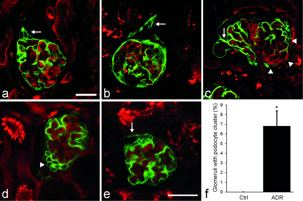
Podocytes are critical in the maintenance of a healthy glomerular filter; however, they have been difficult to study in the intact kidney because of technical limitations. Here we report the development of serial multiphoton microscopy (MPM) of the same glomeruli over several days to visualize the motility of podocytes and parietal epithelial cells (PECs) in vivo. In podocin-GFP mice, podocytes formed sporadic multicellular clusters after unilateral ureteral ligation and migrated into the parietal Bowman's capsule. The tracking of single cells in podocin-confetti mice featuring cell-specific expression of CFP, GFP, YFP or RFP revealed the simultaneous migration of multiple podocytes. In phosphoenolpyruvate carboxykinase (PEPCK)-GFP mice, serial MPM found PEC-to-podocyte migration and nanotubule connections. Our data support a highly dynamic rather than a static nature of the glomerular environment and cellular composition. Future application of this new approach should advance our understanding of the mechanisms of glomerular injury and regeneration.


Conflict of interest statement

Conflict of interest/disclosure

None of the authors has any conflicts of interest to declare.

Figures

Figure 1
Figure 1. MPM imaging in vivo reveals signs of podocyte migration in the intact kidney in the model of unilateral ureteral obstruction (UUO) in Pod-GFP mice
(a) In a wild-type mouse kidney the glomerular podocytes appear as dark, unlabeled cells around capillaries (arrows). Plasma is labeled red with Alexa594-Albumin. Lucifer yellow (yellow) was injected iv to label the filtration (Bowman’s) space. (b) Low-power image of glomeruli after UUO shows restriction of GFP fluorescence to podocytes in Pod-GFP mice. Early podocyte changes that appear after UUO include the development of podocyte cell clusters (c) and their projections protruding (arrow) into the lumen of the proximal tubule (d). (e) Detached, individual podocytes can be seen in the lumen of the proximal tubule. GFP+ cells in the parietal layer appear to develop by visceral podocytes migrating to the parietal layer via the vascular pole transition (f) or directly over the Bowman’s space. (g) Thin connections appear to bridge the parietal and visceral layers of the Bowman’s capsule (arrows). Continuous GFP+ cell coverage in the parietal layer (g) develops later, >3–4 weeks after UUO. Scale bars are 20µm. Magnification is the same on all panels except panel b. (h) Percentage of glomeruli in which >50% of the parietal layer is covered by GFP+ cells in age-matched control mice (n=19) and 2–5 weeks after UUO (n=43). *: P<0.05. Data are presented as mean ± s.e.m. (i) Linear fit of data points (Pod-GFP in black (n=48 mice), iPod-GFP in red (n=5 mice) shows the percentage of glomeruli with >50% parietal GFP+ cell coverage.
Figure 2
Figure 2. Confirmation of podocyte clustering and migration in iPod-GFP mice after UUO (a–c) and podocyte clustering in the model of adriamycin nephropathy in Pod-GFP mice (d–f)
In mice with tamoxifen-induced podocyte GFP expression (iPod-GFP) podocyte clustering was observed <2 weeks after UUO (a, arrow). After >4 weeks following UUO, podocyte projections to the parietal Bowman’s capsule developed either at the urinary pole invading the glomerulo-tubular junction (b, arrow), or at the vascular pole transition (c, arrowheads), or anywhere in between the poles (c, arrow) giving rise to parietal GFP+ cells. Plasma was labeled red with Alexa594-Albumin. MPM imaging of the intact Pod-GFP mouse kidney after adriamycin treatment revealed the presence of podocyte clusters around capillaries (d, arrowhead) and the development of GFP+ cell coverage in the parietal layer which was continuous with the podocyte clusters via the vascular pole transition (e, arrow). Scale bar is 20µm for all panels. (f) Summary of histological analysis showing the percentage of glomeruli with podocyte clusters in age-matched control mice and 4 days after adriamycin treatment (n=4 each). *: P<0.05. Data are presented as mean ± s.e.m.
Figure 3
Figure 3. Serial in vivo MPM imaging of the same glomerulus in Pod-GFP mice after UUO over time, once in 24 hours
(a–b) Podocytes from a hypercellular area at the urinary pole of a collapsed, non-filtering glomerulus (a, arrow) appear to migrate away from the capillary tuft to form a projection into the remainder of the proximal tubule (b, arrow, 24 h later). (c–d) Demonstration of the increased size (growth) of the GFP+ cell projections in the parietal layer (d, arrows) 24 hours after the previous imaging session (c). Plasma is labeled red with Alexa594-Albumin. Scale bar is 20µm for all panels.
Figure 4
Figure 4. Identification and tracking of single podocytes in the multi-color Pod-Confetti mouse model using in vivo MPM imaging
(a) Podocytes are labeled in one of the four colors, either by membrane-targeted CFP, nuclear GFP, cytosolic YFP or cytosolic RFP. Labeled dextran (plasma dye) is shown in grayscale. (b) A CFP-labeled (blue) visceral podocyte is shown bridging over the Bowman’s space to the parietal layer and in direct contact with other parietal cells of the same color (blue arrow). Other adjacent parietal Confetti+ cells are labeled with RFP (red arrow) or GFP (green arrow). (c) Analysis of the Confetti+ cell projections between the visceral and parietal layers shown for both single, isolated projections and for multiple projections in which parietal Confetti+ cells had direct contact with other projections (n=167 single and n=217 multiple projections from 112 glomeruli in 4 mice). (d–e) Serial MPM imaging of the same glomerulus shows the appearance of a nuclear GFP-labeled podocyte around a glomerular capillary (e) compared to 24h prior (d). Arrows indicate the same glomerular tuft region). d–e are projection images of 8 different confocal z-planes. (f) Cell morphology of podocytes including the interdigitating foot processes between two adjacent CFP and YFP-labeled podocytes are visualized in great detail (fixed, frozen tissue section). Scale bar is 20µm for all panels except panel f (1µm).
Figure 5
Figure 5. MPM imaging of PEC migration in vivo in the PEPCK-GFP mouse model after UUO
(a) Cell-specific GFP expression in the proximal tubule and in some, but not all PECs. All other cells are labeled with Tomato. (b–c) Serial MPM imaging of the same glomerulus shows a GFP expressing PEC at the vascular pole of the glomerulus that forms projections propagating into both the visceral and parietal layers. The length of the green projections increased between the time points of 0h (b), and 48 h (c). (d) Four green nanotubules (arrows) are visualized in the intact glomerulus, which appear to connect the PEC layer with the glomerular tuft. Scale bar is 20µm for all panels.

References

    1. Patrakka J, Tryggvason K. New insights into the role of podocytes in proteinuria. Nat Rev Nephrol. 2009;5:463–468. - PubMed
    1. Pavenstädt H, Kriz W, Kretzler M. Cell Biology of the Glomerular Podocyte. Physiol Rev. 2003;83:253–307. - PubMed
    1. Boute N, et al. NPHS2, encoding the glomerular protein podocin, is mutated in autosomal recessive steroid-resistant nephrotic syndrome. Nat Genet. 2000;24:349–354. - PubMed
    1. Huber TB, et al. Bigenic mouse models of focal segmental glomerulosclerosis involving pairwise interaction of CD2AP, Fyn, and synaptopodin. J Clin Invest. 2006;116:1337–1345. - PMC - PubMed
    1. Jones N, et al. Nck adaptor proteins link nephrin to the actin cytoskeleton of kidney podocytes. Nature. 2006;440:818–823. - PubMed

Publication types

MeSH terms

Substances