. 2014 Feb;44(2):505-13.
doi: 10.3892/ijo.2013.2184. Epub 2013 Nov 21.

Hepatic protection and anticancer activity of curcuma: a potential chemopreventive strategy against hepatocellular carcinoma

Affiliations

Hepatic protection and anticancer activity of curcuma: a potential chemopreventive strategy against hepatocellular carcinoma

Yan Li et al. Int J Oncol. 2014 Feb.

Abstract

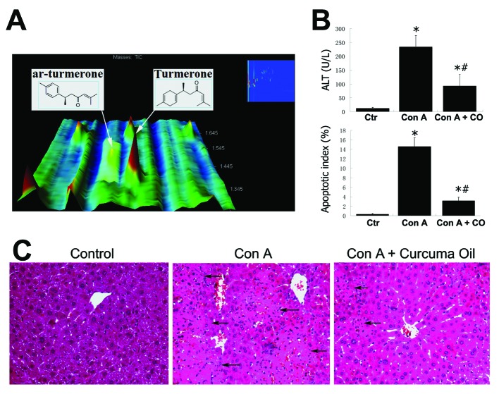
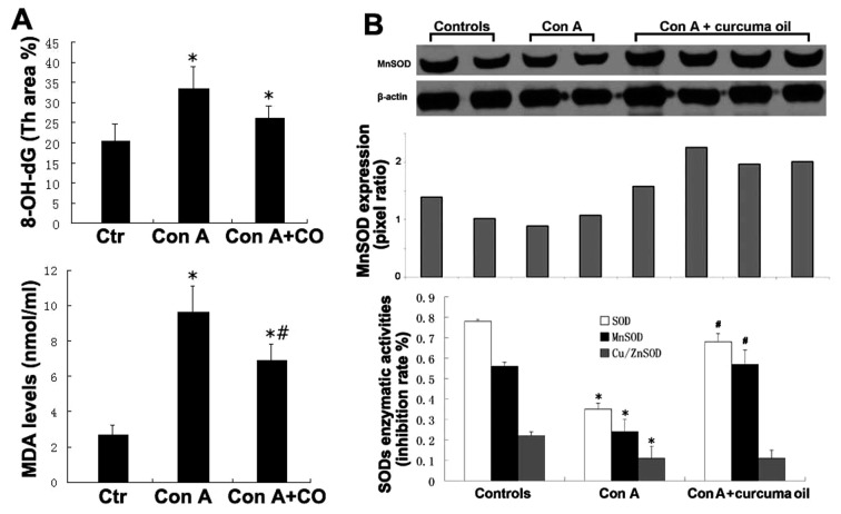
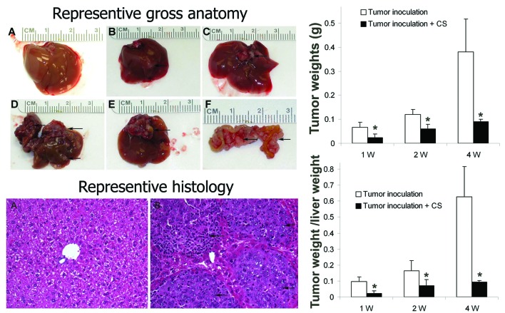
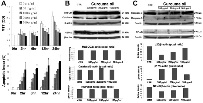
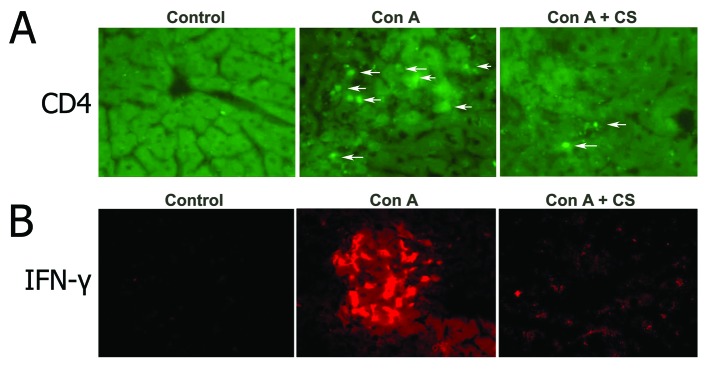
Malignant transformation of hepatocellular carcinoma (HCC) occurs through repetitive liver injury in a context of inflammation and oxidative DNA damage. A spectrum of natural sesquiterpenoids from curcuma oil has displayed antioxidant, anti-inflammatory and anti-carcinogenic properties. The aim of the study was to investigate the hepatoprotective and anti-HCC effects of curcuma oil in vivo and in vitro. Mice were pretreated with curcuma oil (100 mg/kg) for 3 days, then treated with Concanavalin A (30 mg/kg). The hepatic tissue was evaluated for histology, CD4+ cell, interferon-γ, apoptosis, lipid peroxidation, 8-hydroxy-deoxyguanosine and MnSOD. C57L/J mice were treated with curcuma oil and 107 Hepa1-6 cells directly inoculated into liver lobes. The effects of curcuma oil on cell growth and cell death were evaluated. In addition, MnSOD, HSP60, catalase, NF-κB and caspase-3 were also investigated in the Hepa1-6 cells treated with curcuma oil. Pretreatment with curcuma oil significantly attenuates inflammation and oxidative damage by Concanavalin A. Treatment with curcuma oil can decrease the incidence of HCC. Curcuma oil inhibits cell growth and induces cell death in Hepa1-6 cells. Curcuma protected mice with hepatic injury from inflammatory and oxidative stress. Curcuma oil can inhibit hepatoma cell growth in vivo and in vitro.


Figures

Figure 1.
Figure 1.
The potential active ingredients and hepatic protection of curcuma oil. (A) ar-turmerone eluted from the comprehensive GCxGC at 2154 sec in the first dimension column and 1.445 sec in the second dimension column with a peak area of 47342 and a spectrum similarity of 825. α-turmerone was detected at 2164 sec in the first dimension retention time and 1.365 sec in the second dimension retention time with a peak area of 146572 and a spectrum similarity of 859. The spectrum similarity ranges from 0 to 1,000. A high spectrum similarity indicates the high confidence of turmerone identification. (B) The effect of curcuma oil on alanine aminotransferase (ALT) level and apoptosis in hepatic tissue. Ctr, control group; Con A, Con A treated group; Con A + CO, curcuma oil pretreatment + Con A treated group. *p<0.05 vs controls; #p<0.05 vs Con A group. (C) Histological changes in hepatic tissue with Con A challenge. Arrows, necrosis. Hematoxylin and eosin staining (×200).
Figure 2.
Figure 2.
The oxidative damage and hepatic protection of curcuma oil. (A) Effect of curcuma oil on 8-OH-dG and lipid peroxidation in hepatic tissue. 8-OH-dG was detected by immunohistochemistry using computer image-analysis. Th area, the computer program quantified the threshold area represented by color images. Malondialdehyde (MDA) concentrations were measured representing the level of lipid peroxidation. Ctr, control group; Con A, Con A treated group; Con A + CO, curcuma oil pretreatment + Con A treated group. *p<0.05 vs controls. #p<0.05 vs Con A group. (B) The effect of curcuma oil on MnSOD protein expression and enzymatic activity. MnSOD expression was determined by western blotting and the optical density was further quantified by computer imaging software. Pixel ratio (MnSOD/β-actin) was used as the MnSOD expression levels. Enzymatic activities of SODs were determined by a colorimetric assay. SOD, total SOD enzymatic activity; MnSOD, MnSOD enzymatic activity; Cu/ZnSOD; Cu/ZnSOD enzymatic activity. CS, *p<0.05 vs controls. #p<0.05 vs Con A group.
Figure 3.
Figure 3.
Representative figures of liver cancer and tumor weight. Left upper, representative images of gross anatomy of the liver cancer model. (A) Normal liver. (B) Tumor growth in the lobe of inoculation. (C) Tumor cells grow in site other than original inoculation. (D and E) Tumor invaded into the other lobes. (F) Inoculated tumor metastasized into intestinal mesenterium. Left bottom, representative histology of the liver cancer model. (A) Normal liver. (B) Liver cancer. Arrows, inoculated tumor cells. Hematoxylin and eosin staining (×200). Right upper, tumor weights in Hepa1-6 cell inoculation and in Hepa1-6 cell inoculation with pretreatment of curcuma sesquiterpenoids. Right bottom, tumor weight/liver weight in Hepa1-6 cell inoculation and in Hepa1-6 cell inoculation with pretreatment of curcuma sesquiterpenoids. CS, curcuma sesquiterpenoids. W, week. *p<0.05 vs controls.
Figure 4.
Figure 4.
The effects of curcuma oil on hepatoma cells in vitro. (A) Curcuma oil inhibits growth and induces apoptosis in hepatoma cells. Hepa1-6 cells were seeded in a 96-well plate at 2×10⁵ cells/well. Curcuma sesquiterpenoids were added at the concentrations 50, 100, 200 and 500 μg/ml for 2, 6, 12 and 24 h. MTT assay was used to determine cell viability. Spectrophotometric OD value was used as the cell growth index. TUNEL assay was used to determine cell apoptosis. Results are presented as the percentage of apoptotic cells over total cells. The values are expressed as means ± SD. *p<0.05 vs untreated cells (0 μg/ml). (B and C) Effect of curcuma oil on MnSOD, catalase, HSP60, caspase-3 and NF-κB in Hepa1-6 cells. Hepa1-6 cells were seeded in 100-mm dish at 1×10⁶ cells and reached 95% confluence. The cells were treated with curcuma oil at the concentrations 100, 200 and 500 μg/ml for 2 h. After treatment, total protein was extracted to perform the western blotting. The optical density was further quantified by computer imaging software and the pixel ratio was used as the expression levels. CTR, control (untreated).
Figure 5.
Figure 5.
Effect of curcuma sesquiterpenoids on CD4+ T-cells and IFN-γ in hepatic tissue with Con A challenge. (A) FITC-immunofluorescence detection of CD4+ T-cells (×200). (B) Rhodamine-immunofluorescence detection of IFN-γ. Arrows, CD4 positive cells. CS, curcuma sesquiterpenoids.

References

    1. Farazi PA, DePinho RA. Hepatocellular carcinoma pathogenesis: from genes to environment. Nat Rev Cancer. 2006;6:674–687. - PubMed
    1. Levrero M. Viral hepatitis and liver cancer: the case of hepatitis C. Oncogene. 2006;25:3834–3847. - PubMed
    1. Moriya K, Nakagawa K, Santa T, et al. Oxidative stress in the absence of inflammation in a mouse model for hepatitis C virus-associated hepatocarcinogenesis. Cancer Res. 2001;61:4365–4370. - PubMed
    1. Moriya K, Fujie H, Shintani Y, et al. The core protein of hepatitis C virus induces hepatocellular carcinoma in transgenic mice. Nat Med. 1998;4:1065–1067. - PubMed
    1. Koike K, Tsutsumi T, Miyoshi H, et al. Molecular basis for the synergy between alcohol and hepatitis C virus in hepatocarcinogenesis. J Gastroenterol Hepatol 23. 2008;(Suppl 1):S87–S91. - PubMed

Publication types

MeSH terms