

Estimating the size and number of autophagic bodies by electron microscopy
- PMID: 24270884
- PMCID: PMC4389869
- DOI: 10.4161/auto.26856
Estimating the size and number of autophagic bodies by electron microscopy
Abstract
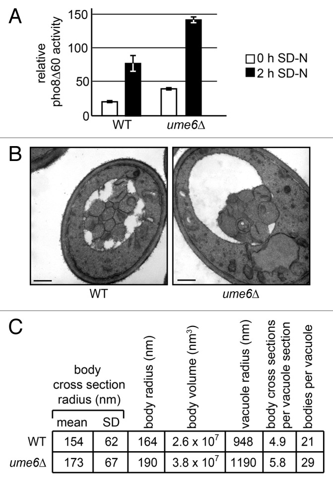
Much recent and ongoing research is focused on understanding the mechanisms and regulation of autophagy, a cellular self-degradation pathway with many links to human health. Although many assays exist to measure the total magnitude of autophagy, electron microscopy remains the tool of choice for the determination of the size and the number of autophagosomes formed in a given mutant or under given induction conditions. Here we present a detailed protocol for measuring autophagic bodies in the yeast Saccharomyces cerevisiae by electron microscopy. Furthermore, we present an improved mathematical method for estimating body size and a new method for estimating body number. Finally, we include a discussion of the merits and limitations of these methods and an example of their application to autophagic bodies formed in the ume6∆ strain.
Keywords: autophagosome; autophagy; electron microscopy; stereology; vacuole; yeast.
Figures
References
-
- Xie Z, Nair U, Geng J, Szefler MB, Rothman ED, Klionsky DJ. . Indirect estimation of the area density of Atg8 on the phagophore. Autophagy 2009; 5:217 - 20; http://dx.doi.org/ 10.4161/auto.5.2.7201; PMID: 19088501 - DOI - PMC - PubMed
-
- Levine B, Kroemer G. . Autophagy in the pathogenesis of disease. Cell 2008; 132:27 - 42; http://dx.doi.org/ 10.1016/j.cell.2007.12.018; PMID: 18191218 - DOI - PMC - PubMed
-
- Klionsky DJ. . Autophagy: from phenomenology to molecular understanding in less than a decade. Nat Rev Mol Cell Biol 2007; 8:931 - 7; http://dx.doi.org/ 10.1038/nrm2245; PMID: 17712358 - DOI - PubMed
-
- Knorr RL, Dimova R, Lipowsky R. . Curvature of double-membrane organelles generated by changes in membrane size and composition. PLoS One 2012; 7:e32753; http://dx.doi.org/ 10.1371/journal.pone.0032753; PMID: 22427874 - DOI - PMC - PubMed
-
- Xie Z, Nair U, Klionsky DJ. . Atg8 controls phagophore expansion during autophagosome formation. Mol Biol Cell 2008; 19:3290 - 8; http://dx.doi.org/ 10.1091/mbc.E07-12-1292; PMID: 18508918 - DOI - PMC - PubMed
Publication types
MeSH terms
Grants and funding
LinkOut - more resources
Full Text Sources
Other Literature Sources
Molecular Biology Databases