. 2013 Nov;46(11):936-942.
doi: 10.1590/1414-431X20132764. Epub 2013 Nov 6.

Performance assessment of the single photon emission microscope: high spatial resolution SPECT imaging of small animal organs

Affiliations

Performance assessment of the single photon emission microscope: high spatial resolution SPECT imaging of small animal organs

J Mejia et al. Braz J Med Biol Res. 2013 Nov.

Abstract

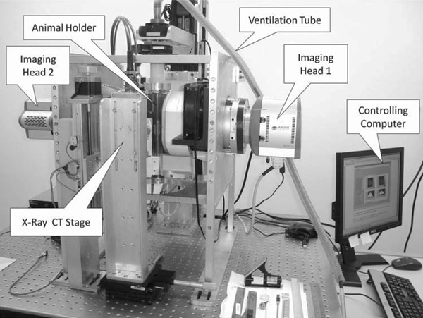
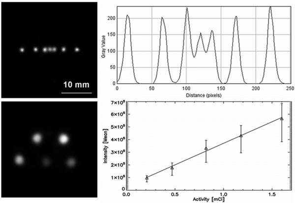
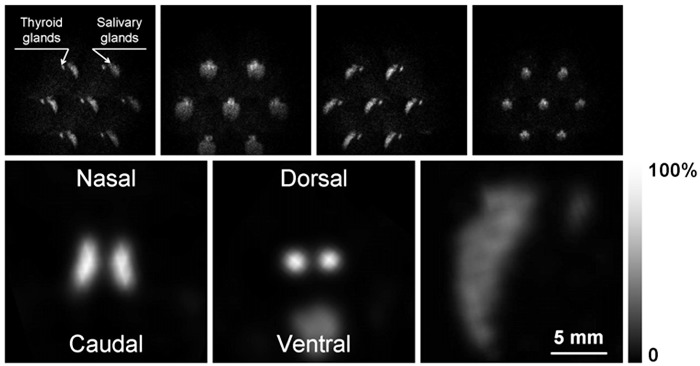
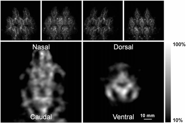
The single photon emission microscope (SPEM) is an instrument developed to obtain high spatial resolution single photon emission computed tomography (SPECT) images of small structures inside the mouse brain. SPEM consists of two independent imaging devices, which combine a multipinhole collimator, a high-resolution, thallium-doped cesium iodide [CsI(Tl)] columnar scintillator, a demagnifying/intensifier tube, and an electron-multiplying charge-coupling device (CCD). Collimators have 300- and 450-µm diameter pinholes on tungsten slabs, in hexagonal arrays of 19 and 7 holes. Projection data are acquired in a photon-counting strategy, where CCD frames are stored at 50 frames per second, with a radius of rotation of 35 mm and magnification factor of one. The image reconstruction software tool is based on the maximum likelihood algorithm. Our aim was to evaluate the spatial resolution and sensitivity attainable with the seven-pinhole imaging device, together with the linearity for quantification on the tomographic images, and to test the instrument in obtaining tomographic images of different mouse organs. A spatial resolution better than 500 µm and a sensitivity of 21.6 counts·s-1·MBq-1 were reached, as well as a correlation coefficient between activity and intensity better than 0.99, when imaging 99mTc sources. Images of the thyroid, heart, lungs, and bones of mice were registered using 99mTc-labeled radiopharmaceuticals in times appropriate for routine preclinical experimentation of <1 h per projection data set. Detailed experimental protocols and images of the aforementioned organs are shown. We plan to extend the instrument's field of view to fix larger animals and to combine data from both detectors to reduce the acquisition time or applied activity.


Figures

Figure 1
Figure 1. Single photon emission microscope (SPEM). The main components are labeled.
Figure 2
Figure 2. Spatial resolution and quantification. Upper panels, capillary tube phantom. Left: characteristic slice through the volumetric reconstruction of the phantom's emission. Right: intensity profile along a line intersecting the seven capillaries. It can be seen that all the capillary's emissions are well identified. Lower panels, quantification phantom. Left: characteristic slice through the volumetric reconstruction of the phantom's emission. Right: plot of the activity in the phantom versus mean intensity in the phantom.
Figure 3
Figure 3. Thyroid images. Upper panels, sequence of 4 of the 16 projections obtained with the seven-pinhole imaging device of the mouse neck area, labeled with sodium pertechnetate. Lower panels, characteristic slices through the reconstructed volumetric model of the radioactive emission from the mouse thyroid and salivary glands along the three main axes, after 80 iterations.
Figure 4
Figure 4. Cardiac images. Characteristic slices perpendicular to the main axis of the reconstructed volumetric model of the radioactive emission from the mouse heart, labeled with 99mTc-MIBI, after 80 iterations.
Figure 5
Figure 5. Pulmonary images. Upper panels, sequence of 4 of the 16 projections obtained with the seven-pinhole imaging device of the mouse chest area, labeled with 99mTc-MAA. Lower panels, characteristic transversal and coronal slices through the reconstructed volumetric model of the radioactive emission from the mouse lungs, after 60 iterations.
Figure 6
Figure 6. Bone images. Upper panels, sequence of 4 of the 16 projections obtained with the seven-pinhole imaging device of the mouse head area, labeled with 99mTc-MDP. Lower panels, characteristic transversal and coronal slices through the reconstructed volumetric model of the radioactive emission from the mouse skull, after 80 iterations.

Similar articles

Cited by

References

    1. Beekman F, van der Have F. The pinhole: gateway to ultra-high-resolution three-dimensional radionuclide imaging. Eur J Nucl Med Mol Imaging. 2007;34:151–161. doi: 10.1007/s00259-006-0248-6. - DOI - PubMed
    1. Golestani R, Wu C, Tio RA, Zeebregts CJ, Petrov AD, Beekman FJ, et al. Small-animal SPECT and SPECT/CT: application in cardiovascular research. Eur J Nucl Med Mol Imaging. 2010;37:1766–1777. doi: 10.1007/s00259-009-1321-8. - DOI - PMC - PubMed
    1. Mejia J, Galvis-Alonso OY, Castro AA, Braga J, Leite JP, Simoes MV. A clinical gamma camera-based pinhole collimated system for high resolution small animal SPECT imaging. Braz J Med Biol Res. 2010;43:1160–1166. doi: 10.1590/S0100-879X2010007500128. - DOI - PubMed
    1. Difilippo FP. Design and performance of a multi-pinhole collimation device for small animal imaging with clinical SPECT and SPECT-CT scanners. Phys Med Biol. 2008;53:4185–4201. doi: 10.1088/0031-9155/53/15/012. - DOI - PMC - PubMed
    1. Wagenaar DJ, Engdahl JC, Simcic V, Hawman EG, Mertelmeier T, Mahmood U, et al. Use of conventional gamma cameras for small animal imaging. IEEE Nucl Sci Symp Conf Rec. 2000:86–90.