. 2014 Feb;42(4):e29.
doi: 10.1093/nar/gkt1179. Epub 2013 Nov 22.

Highly specific and sensitive method for measuring nucleotide excision repair kinetics of ultraviolet photoproducts in human cells

Affiliations

Highly specific and sensitive method for measuring nucleotide excision repair kinetics of ultraviolet photoproducts in human cells

Jun-Hyuk Choi et al. Nucleic Acids Res. 2014 Feb.

Abstract

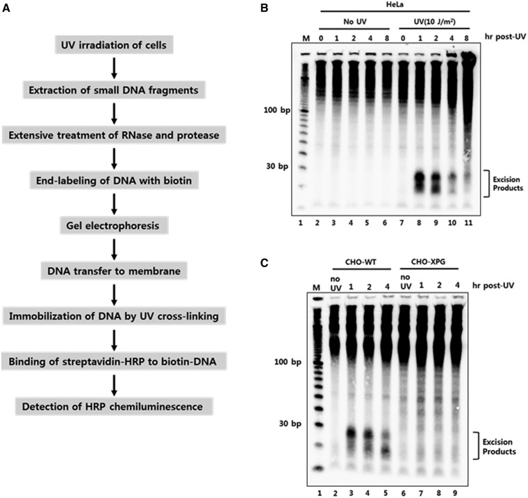
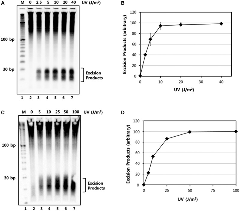
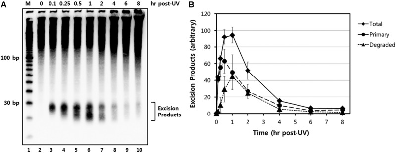
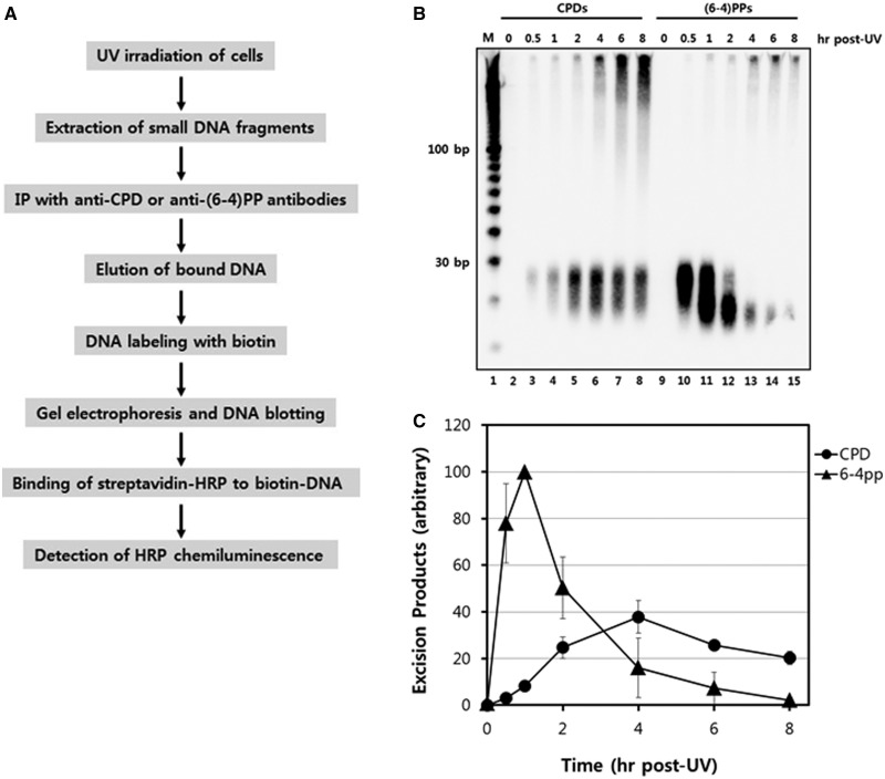
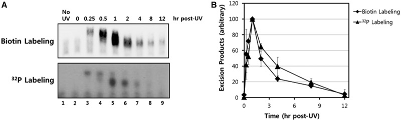
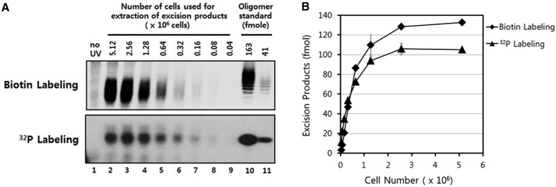
The nucleotide excision repair pathway removes ultraviolet (UV) photoproducts from the human genome in the form of short oligonucleotides ∼ 30 nt in length. Because there are limitations to many of the currently available methods for investigating UV photoproduct repair in vivo, we developed a convenient non-radioisotopic method to directly detect DNA excision repair events in human cells. The approach involves extraction of oligonucleotides from UV-irradiated cells, DNA end-labeling with biotin and streptavidin-mediated chemiluminescent detection of the excised UV photoproduct-containing oligonucleotides that are released from the genome during excision repair. Our novel approach is robust, with essentially no signal in the absence of UV or a functional excision repair system. Furthermore, our non-radioisotopic methodology allows for the sensitive detection of excision products within minutes following UV irradiation and does not require additional enrichment steps such as immunoprecipitation. Finally, this technique allows for quantitative measurements of excision repair in human cells. We suggest that the new techniques presented here will be a useful and powerful approach for studying the mechanism of human nucleotide excision repair in vivo.


Figures

Figure 1.
Figure 1.
Development of a novel non-radioisotopic method for the detection of excised oligonucleotide products of nucleotide excision repair in vivo. (A) Schematic representation for the detection of excised oligonucleotides containing UV damage in human cells. UV-irradiated cells are harvested and subjected to DNA extraction to isolate low molecular weight DNAs. After extensive treatment with RNase and protease, the short DNA molecules are labeled with biotin at the 3′-end using terminal deoxynucleotidyl transferase (TdT). The biotin-labeled oligonucleotides are separated by gel electrophoresis and then transferred to a nylon membrane. The transferred biotin-DNA molecules are immobilized by UV cross-linking, incubated with HRP-conjugated streptavidin, and detected with a chemiluminescence reagent. (B) HeLa cells were exposed to 10 J/m2 of UV-C and then harvested at different time points. The cells were subjected to the modified Hirt lysis and labeling procedures as in A. The DNA marker (M) is a 10 bp DNA ladder that was labeled with biotin and loaded onto the gel. (C) CHO wild-type (AA8) and XPG-deficient (UV135) cells were harvested at different times after UV exposure to 10 J/m2 of UV-C. Excised oligonucleotides containing UV damage were then extracted and processed as described in A.
Figure 2.
Figure 2.
UV dose-dependent generation of excised oligonucleotides. (A) HeLa cells were treated with the indicated doses of UV-C radiation and then harvested after incubation for 30 min. Excised oligonucleotides containing UV damage were then extracted, 3′ end-labeled with biotin, separated on a 10% TBE-urea gel, transferred to a nylon membrane, and detected with streptavidin-HRP conjugate using chemiluminescence reagents. (B) Quantitative analysis of the excision repair products from three independent experiments as in A are presented as means ± standard deviation (SD). The maximum values were set to 100 and the other values are presented relative to that value. (C) A375 cells were treated with the indicated doses of UV-C radiation and then harvested 1 h later. Excised oligonucleotides were analyzed as in A, except with a 12% TBE-urea gel. (D) Quantitative analysis of the excised oligonucleotide repair products in A375 cells.
Figure 3.
Figure 3.
Time course analysis of UV-induced excision products. (A) HeLa cells were exposed to 10 J/m2 of UV-C and then harvested at various time points. Excised oligonucleotides containing UV damage were then extracted, 3′ end-labeled with biotin, separated on a 10% TBE-urea gel, transferred to a nylon membrane, and detected with HRP-conjugated streptavidin and a chemiluminescent reagent. (B) Quantitative analysis of the excised oligonucleotide repair products. The results from three independent experiments were quantified and are plotted (means ± standard deviation) relative to the maximum.
Figure 4.
Figure 4.
Detection of specific excised oligonucleotides containing CPDs or (6–4)PPs. (A) Scheme for the detection of excised oligonucleotides containing UV photoproduct. (B) HeLa cells were harvested at various time points after UV exposure to 10 J/m2 of UV-C. The cells were subjected to the modified Hirt procedure, and the extracted oligonucleotides were then immunoprecipitated with anti-(6–4)PPs or anti-CPD antibodies. Purified oligonucleotides 3′-end labeled with biotin-11-UTP were separated on a 10% TBE-urea gel, transferred to a nylon membrane, and detected with HRP-conjugated streptavidin using a chemiluminescent reagent. (C) Quantitative analysis of the CPD- and (6–4)PP-containing excised oligonucleotides products as a function of time following UV irradiation. The results from three independent experiments were quantified and are plotted (means ± standard deviation) relative to the maximum.
Figure 5.
Figure 5.
Sensitivity comparison of the biotinylation and radiolabeling methods. (A) Excised oligonucleotides containing UV damage were recovered from A375 cells at various time points after exposure to 10 J/m2 of UV-C. The purified oligonucleotides were then labeled with biotin-11-dUTP or cordycepin 5′-triphosphate at the 3′-end using terminal deoxynucleotidyl transferase (TdT). Biotin-labeled DNAs were separated on a 12% TBE-urea gel, transferred to a nylon membrane, and detected with HRP-conjugated streptavidin-HRP using a chemiluminescence reagent. Radiolabeled DNAs were separated on a 12% TBE-urea gel and detected by exposing the gel to a phosphorimager screen. (B) Quantitative analysis of the excised repair products. The results from three independent experiments were quantified and are plotted (means ± standard deviation) relative to the maximum.
Figure 6.
Figure 6.
Absolute quantification of UV photoproducts in vivo. (A) Excised oligonucleotides containing UV damage were recovered from A375 cells 1 h following exposure to 10 J/m2 of UV-C. The purified oligonucleotides were then either biotinylated or radiolabeled as in Figure 5 using Hirt lysate-purified DNA from a defined number of cells. As a standard for quantification of the excised oligonucleotides, a 20-nt long single-stranded DNA oligonucleotide was either biotinylated or radiolabeled and then electrophoresed and analyzed with the UV photoproduct-containing excised oligomer repair products. (B) Absolute quantification of the excised repair products in vivo. The results from two independent experiments were quantified and are plotted (means ± standard deviation).

References

    1. Sancar A. DNA excision repair. Annu. Rev. Biochem. 1996;65:43–81. - PubMed
    1. Wood RD. Nucleotide excision repair in mammalian cells. J. Biol. Chem. 1997;272:23465–23468. - PubMed
    1. Reardon JT, Sancar A. Nucleotide excision repair. Prog. Nucleic Acid Res. Mol. Biol. 2005;79:183–235. - PubMed
    1. Huang JC, Svoboda DL, Reardon JT, Sancar A. Human nucleotide excision nuclease removes thymine dimers from DNA by incising the 22nd phosphodiester bond 5' and the 6th phosphodiester bond 3' to the photodimer. Proc. Natl Acad. Sci. USA. 1992;89:3664–3668. - PMC - PubMed
    1. Svoboda DL, Taylor JS, Hearst JE, Sancar A. DNA repair by eukaryotic nucleotide excision nuclease. Removal of thymine dimer and psoralen monoadduct by HeLa cell-free extract and of thymine dimer by xenopus laevis oocytes. J. Biol. Chem. 1993;268:1931–1936. - PubMed

Publication types