Review
. 2014 Feb;7(2):181-92.
doi: 10.1242/dmm.012245. Epub 2013 Nov 21.

Beyond the zebrafish: diverse fish species for modeling human disease

Affiliations
Review

Beyond the zebrafish: diverse fish species for modeling human disease

Manfred Schartl. Dis Model Mech. 2014 Feb.

Abstract

In recent years, zebrafish, and to a lesser extent medaka, have become widely used small animal models for human diseases. These organisms have convincingly demonstrated the usefulness of fish for improving our understanding of the molecular and cellular mechanisms leading to pathological conditions, and for the development of new diagnostic and therapeutic tools. Despite the usefulness of zebrafish and medaka in the investigation of a wide spectrum of traits, there is evidence to suggest that other fish species could be better suited for more targeted questions. With the emergence of new, improved sequencing technologies that enable genomic resources to be generated with increasing efficiency and speed, the potential of non-mainstream fish species as disease models can now be explored. A key feature of these fish species is that the pathological condition that they model is often related to specific evolutionary adaptations. By exploring these adaptations, new disease-causing and disease-modifier genes might be identified; thus, diverse fish species could be exploited to better understand the complexity of disease processes. In addition, non-mainstream fish models could allow us to study the impact of environmental factors, as well as genetic variation, on complex disease phenotypes. This Review will discuss the opportunities that such fish models offer for current and future biomedical research.

Keywords: Cancer; Evolutionary mutant model; Fish model; Natural variation.


Figures

Fig. 1.
Fig. 1.
Sequence comparison of the human and fish (medaka) HRAS proteins. Below the alignment, identical amino acids are represented by an asterisk (*), a semi-conservative change is indicated by a dot (.) and a conservative change is indicated by a colon (:). The most frequent mutations in the STPase enzymatic active site of the protein in human cancers are found at amino acid positions 12 and 61.
Fig. 2.
Fig. 2.
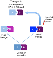
Schematic representation of the process of protein divergence in different lineages after separation from a common ancestor. Because interacting proteins evolve independently, the molecular structure of the interaction interface changes in both partners so that they still fit (i.e. coevolution occurs), but this will not necessarily affect the same amino acids. Eventually, protein A from one lineage is unable to properly interact with protein B from the other lineage (despite having fully retained its biochemical function, e.g. as an enzyme or G-protein). This process is known to evolutionary biologists as the Muller-Dobshansky-Bateson phenomenon. The divergence of interacting proteins can have an impact on studying the effect of a human-derived transgene in the non-human lineage.
Fig. 3.
Fig. 3.
Representative Antarctic notothenioid fish. The Antarctic rockcod, Notothenia coriiceps (top), is red-blooded and possesses a robustly mineralized skeleton. The white-blooded icefish, Chaenocephalus aceratus (bottom), is profoundly anemic and osteopenic. Photographs provided by H. William Detrich (Northeastern University).
Fig. 4.
Fig. 4.
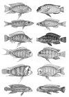
Cichlid fish have evolved different craniofacial morphologies according to the diet to which the respective species has specialized. This has led to differences in the morphology of the skull, and particularly the jaws. Interestingly, similar ecological adaptation has repeatedly led to the evolution of similar morphologies in different cichlid lineages. The picture shows similar ecomorphs from Lake Tanganyika (left) and Lake Malawi (right). Reproduced with permission (Albertson and Kocher, 2006), and outside the scope of the CC-BY license.
Fig. 5.
Fig. 5.
Two epigean Mexican tetras (Astyanax mexicanus) and their eyeless unpigmented cave-dwelling relative. Photo provided by Richard Borowsky (New York University).
Fig. 6.
Fig. 6.
Development of melanoma in hybrid offspring of platyfish and swordtails. (A–C) Platyfish (Xiphophorus maculatus) with different pigmentation patterns are shown. (D,E) After crossing with swordtails (X. hellerii), offspring of A and B develop malignant melanoma. (A) This fish has pigment spots in the dorsal fin; (D) in the hybrids, which carry the Sd (spotted dorsal) allele of Tu and a closely linked tumor modifier, mdl, which determine the onset and compartment of pigment spot or melanoma formation, respectively, melanoma formation spreads out from this compartment. (B) This platyfish has black spots on the flanks determined by the Sp (spotted) allele of Tu and its linked mdl; (E) hybrids consequently develop melanoma from the body sides. Note that the dorsal fin of this fish is free of melanoma cells, which is different from the fish in D. (C,F) The fish in C carries the Sr (striped) allele of Tu along with mdl, which are not tumorigenic in the hybrids (F). The molecular basis of this is so far unknown.
Fig. 7.
Fig. 7.
Healthy and diseased bicolor damselfish (Stegastes partitus). (A) Healthy; (B) diseased. The diseased fish shows several pigmented lesions (chromatophoromas) and a non-pigmented neurofibroma in the corner of the mouth. Photos provided by Michael Schmale (University of Miami).

References

    1. Albertson R. C., Kocher T. D. (2006). Genetic and developmental basis of cichlid trophic diversity. Heredity 97, 211–221 - PubMed
    1. Albertson R. C., Cresko W., Detrich H. W., III, Postlethwait J. H. (2009). Evolutionary mutant models for human disease. Trends Genet. 25, 74–81 - PMC - PubMed
    1. Albertson R. C., Yan Y. L., Titus T. A., Pisano E., Vacchi M., Yelick P. C., Detrich H. W., III, Postlethwait J. H. (2010). Molecular pedomorphism underlies craniofacial skeletal evolution in Antarctic notothenioid fishes. BMC Evol. Biol. 10, 4. - PMC - PubMed
    1. Amatruda J. F., Patton E. E. (2008). Genetic models of cancer in zebrafish. Int Rev Cell Mol Biol 271, 1–34 - PubMed
    1. Amores A., Force A., Yan Y. L., Joly L., Amemiya C., Fritz A., Ho R. K., Langeland J., Prince V., Wang Y. L., et al. (1998). Zebrafish hox clusters and vertebrate genome evolution. Science 282, 1711–1714 - PubMed

Publication types