. 2014 Jan 3;289(1):387-402.
doi: 10.1074/jbc.M113.504779. Epub 2013 Nov 22.

Latrophilins function as heterophilic cell-adhesion molecules by binding to teneurins: regulation by alternative splicing

Affiliations

Latrophilins function as heterophilic cell-adhesion molecules by binding to teneurins: regulation by alternative splicing

Antony A Boucard et al. J Biol Chem. .

Abstract

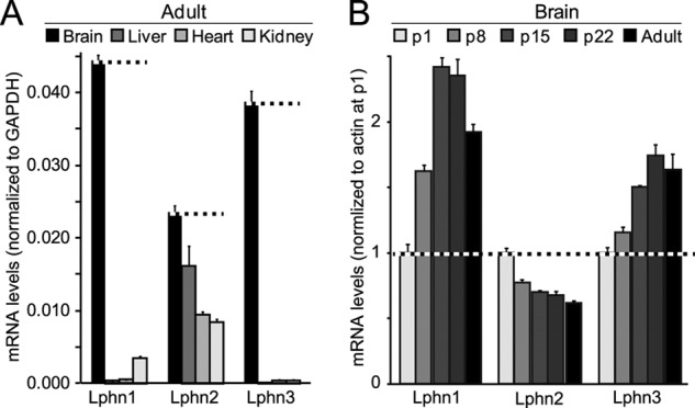
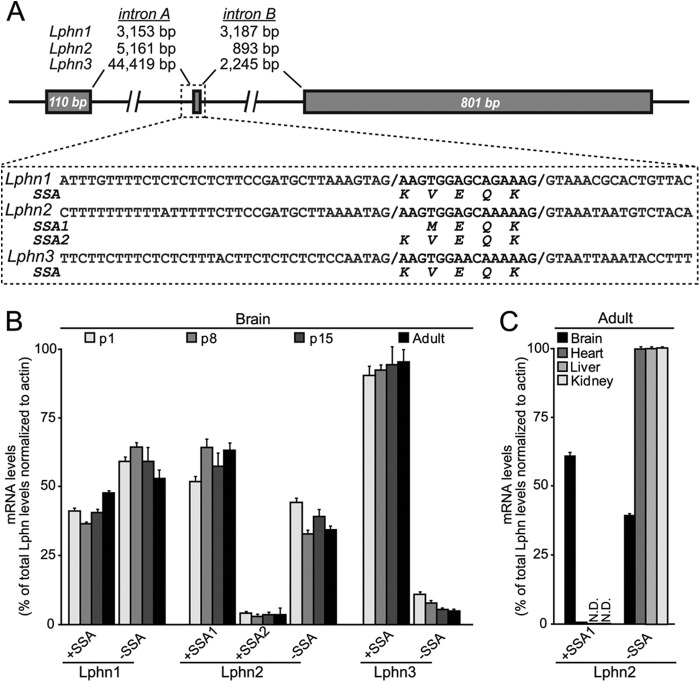
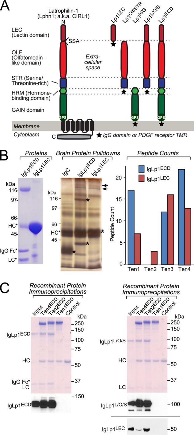
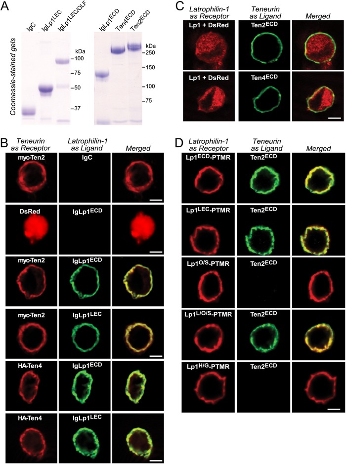
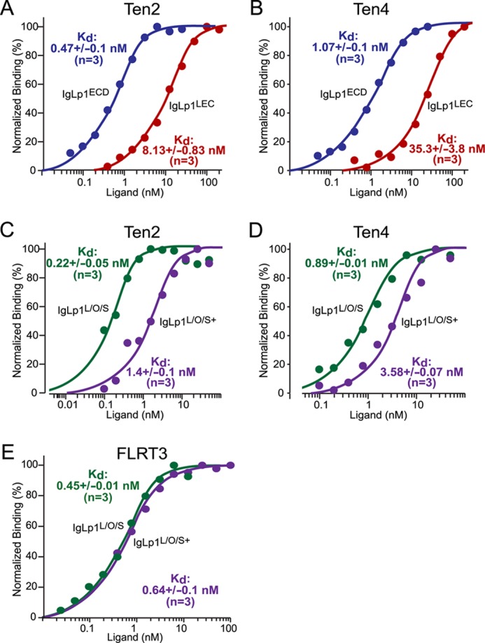
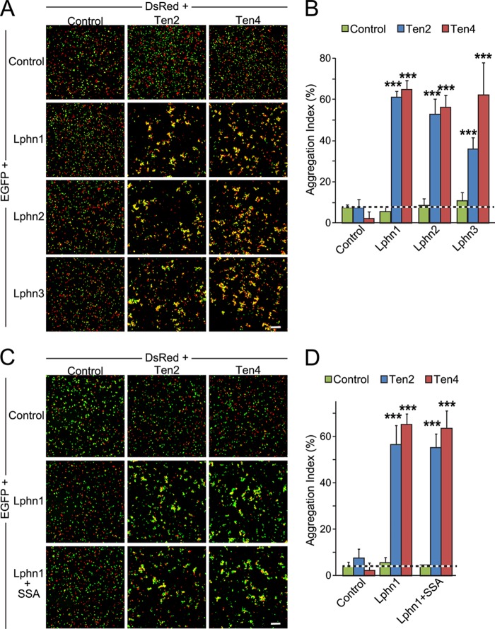
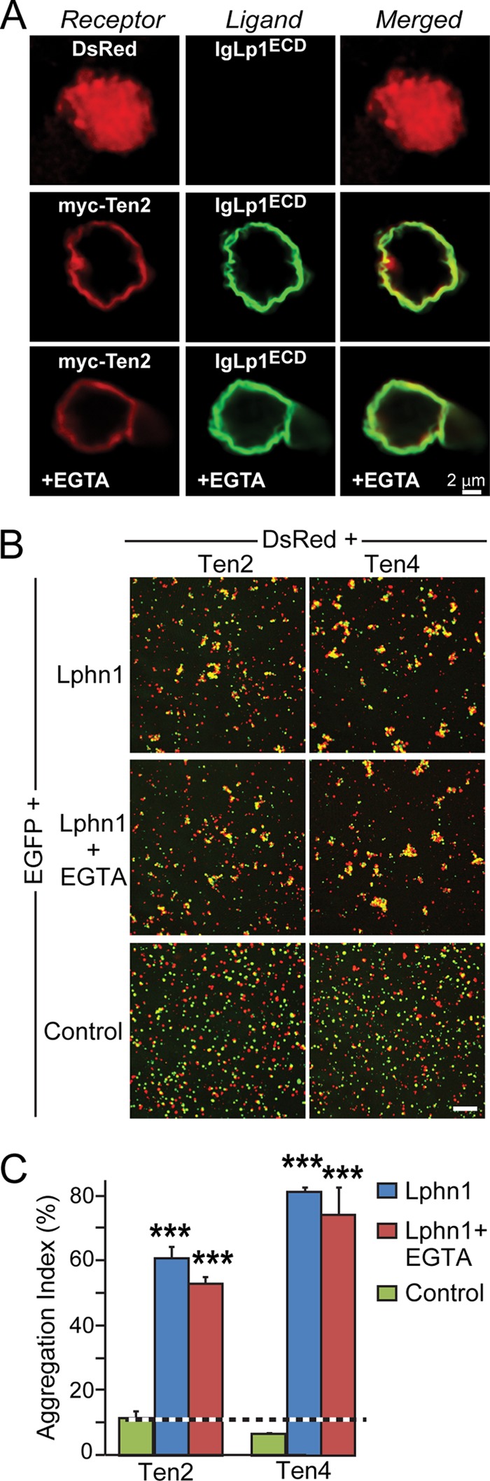
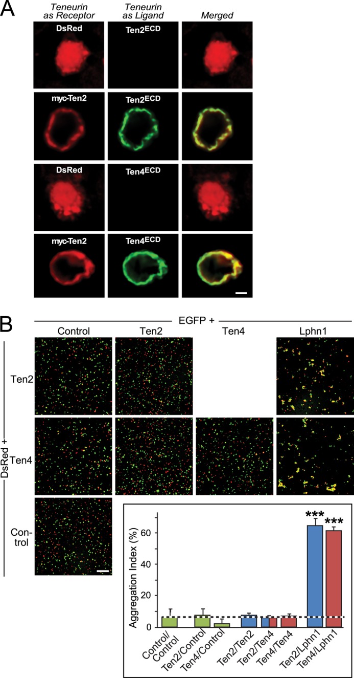
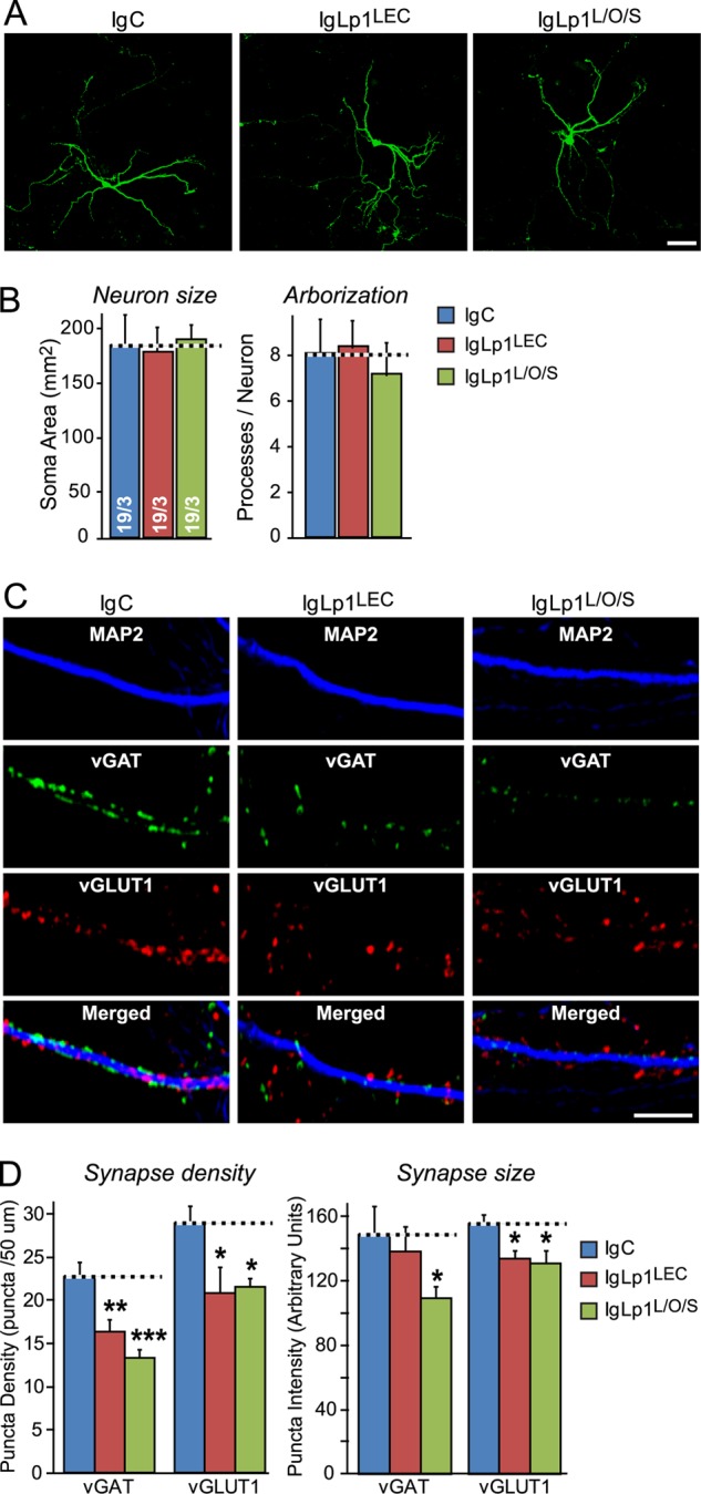
Latrophilin-1, -2, and -3 are adhesion-type G protein-coupled receptors that are auxiliary α-latrotoxin receptors, suggesting that they may have a synaptic function. Using pulldowns, we here identify teneurins, type II transmembrane proteins that are also candidate synaptic cell-adhesion molecules, as interactors for the lectin-like domain of latrophilins. We show that teneurin binds to latrophilins with nanomolar affinity and that this binding mediates cell adhesion, consistent with a role of teneurin binding to latrophilins in trans-synaptic interactions. All latrophilins are subject to alternative splicing at an N-terminal site; in latrophilin-1, this alternative splicing modulates teneurin binding but has no effect on binding of latrophilin-1 to another ligand, FLRT3. Addition to cultured neurons of soluble teneurin-binding fragments of latrophilin-1 decreased synapse density, suggesting that latrophilin binding to teneurin may directly or indirectly influence synapse formation and/or maintenance. These observations are potentially intriguing in view of the proposed role for Drosophila teneurins in determining synapse specificity. However, teneurins in Drosophila were suggested to act as homophilic cell-adhesion molecules, whereas our findings suggest a heterophilic interaction mechanism. Thus, we tested whether mammalian teneurins also are homophilic cell-adhesion molecules, in addition to binding to latrophilins as heterophilic cell-adhesion molecules. Strikingly, we find that although teneurins bind to each other in solution, homophilic teneurin-teneurin binding is unable to support stable cell adhesion, different from heterophilic teneurin-latrophilin binding. Thus, mammalian teneurins act as heterophilic cell-adhesion molecules that may be involved in trans-neuronal interaction processes such as synapse formation or maintenance.

Keywords: Alternative Splicing; Cell Adhesion; FLRT3; G Protein-coupled Receptors (GPCR); Ligand-binding Protein; Neurexin; Neurons; Synapse Formation; Teneurin; α-Latrotoxin.


Figures

FIGURE 1.
FIGURE 1.
Latrophilin-1 and -3 (Lphn1 and Lphn3) are primarily expressed in brain in a developmentally regulated manner, whereas latrophilin-2 (Lphn2) is ubiquitously expressed. A, quantitative RT-PCR measurements of the latrophilin-1, -2, and -3 (Lphn1, Lphn2, and Lphn3) mRNAs in the brain, liver, heart, and kidney of adult mice. mRNA levels were normalized to those of GAPDH monitored in the same samples. B, quantitative RT-PCR measurements of the Lphn1, Lphn2, and Lphn3 mRNAs in brain at postnatal days 1 (p1), 8 (p8), 15 (p15), 22 (p22), and in adult brain. All mRNA levels were normalized to those of actin as an internal control and then to the mRNA levels of the respective latrophilin at p1. Data shown are means ± S.E. (n = 4).
FIGURE 2.
FIGURE 2.
All latrophilin genes contain an alternatively spliced 5′ exon. A, gene structures and sequences flanking the alternatively spliced 5′ exon of latrophilin genes. B, mRNA measurements of alternatively spliced variants of Lphn1, Lphn2, and Lphn3. C, tissue distribution of alternatively spliced Lphn2 variants.
FIGURE 3.
FIGURE 3.
Affinity purification of teneurins with the lectin domain of latrophilin-1. A, domain structure of Lphn1 (left) and of Lphn1 constructs used in this study (right). SSA in Lphn1 contains or lacks a five-residue sequence (KVEQK, after Tyr131). The GAIN domain includes a C-terminal GPS sequence that determines the site of cleavage of the autocatalytic GAIN domain (12). The diagram on the right shows the structure of different Lphn1 fragments that were used for experiments either as purified secreted Ig domain fusion proteins or as surface-displayed fusion proteins with a C-terminal TMR from the PDGF receptor. B, purified recombinant Ig domain fusion proteins of the entire extracellular sequence (IgLp1ECD) or the lectin domain of Lphn1 (IgLp1LEC; see left panel for Coomassie-stained SDS gel of purified proteins) were used for affinity chromatography experiments with total solubilized rat brain proteins, using IgC as a negative control. Bound proteins were eluted with 1.0 m NaCl and analyzed by SDS-PAGE and silver staining (middle panel; asterisks mark eluted bait proteins.). Mass spectrometry of eluted proteins identified fragments of all four teneurin isoforms (right panel). C, pulldown of soluble recombinant Ig domain fusion proteins of the indicated Lphn1 fragments (see A) with immobilized teneurin fragments containing the entire extracellular sequences of teneurin-1 (Ten1ECD), teneurin-2 (Ten2ECD), and teneurin-4 (Ten4ECD). FLAG-tagged teneurins were immobilized on an anti-FLAG antibody column and incubated with the indicated Ig fusion proteins. Beads were washed with incubation buffer, eluted with sample buffer, run on an SDS-polyacrylamide gel, and either stained with Coomassie Blue or analyzed by immunoblotting using an anti-human IgG antibody (to visualize the Ig domain fusion protein) coupled to horseradish peroxidase. Data shown are representative images of experiments that were repeated at least three times.
FIGURE 4.
FIGURE 4.
Surface binding assays reveal domain-specific interactions between Lphn1 and Ten2 and Ten4. A, soluble IgC, Ig-Lp1ECD, IgLp1L/O/S, and IgLp1LEC were produced in transfected HEK293T cells and analyzed by SDS-PAGE and Coomassie Blue staining. B–D, cell surface labeling assays. Transfected HEK293T cells co-expressing membrane-anchored teneurin or latrophilin “receptor” with DsRed were incubated with control medium or medium containing soluble latrophilin Ig domain fusion proteins or soluble teneurin fragments as ligands. Receptors and bound ligands were visualized by immunocytochemistry except for C in which the Lphn1 receptor lacked a tag, and cells were visualized using co-expressed DsRed. B analyzes the binding of latrophilin-1 Ig fusion proteins containing the complete extracellular domains (IgLp1ECD) or only the lectin domain (IgLp1LEC) of Lphn1 to cells expressing DsRed, Myc-tagged Ten2, or HA-tagged Ten4. C examines the binding of soluble Myc-tagged Ten2 and HA-tagged Ten4 proteins containing their entire extracellular domains to cells expressing full-length Lphn1. D monitors the binding of recombinant teneurin proteins to cells expressing latrophilin-1 fusion proteins in which various extracellular latrophilin domains are fused C-terminally to the TMR of the PDGF receptor. The various Lphn1 proteins in D contained the following domains: Lp1ECD-PTMR, the entire extracellular domains; Lp1LEC-PTMR, the lectin domain only; Lp1O/S-PTMR, the olfactomedin-like and serine/threonine-rich sequence; Lp1L/O/S-PTMR, the N-terminal lectin-olfactomedin-like, and serine-rich sequences, and Lp1H/G-PTMR, the hormone binding and GAIN domains. Data shown are from a representative experiment that was independently repeated at least three times.
FIGURE 5.
FIGURE 5.
Binding affinity of Lphn1 to Ten2 and Ten4 and to FLRT3 and effect of alternative splicing of latrophilin-1 on such binding. HEK293T cells expressing Ten2, Ten4, or FLRT3 were used to measure the binding affinity of Ig fusion proteins containing the indicated extracellular Lphn1 domains. All data shown are binding after subtraction of the signal obtained with mock-transfected cells included in all assays. Binding data shown are representative graphs of experiments that were conducted at least three times. Data were fit to a Scatchard equation using SigmaPlot software and assuming uniform binding sites; Kd numbers displayed in the graphs show the averages determined in at least three independent experiments. A–E, binding data for the entire Lphn1 extracellular domains (IgLp1ECD; A and B), the N-terminal Lphn1 lectin domain only (IgLp1LEC; A and B), or the N-terminal three Lphn1 domains (i.e. its lectin, olfactomedin-like, and serine/threonine-rich domains); for the latter, splice variants lacking an insert in SSA (IgLp1L/O/S; C–E) or containing such insert (IgLp1L/O/S+; C–E) were analyzed.
FIGURE 6.
FIGURE 6.
Binding of latrophilins to teneurins mediates cell-cell adhesion. A, HEK293T cells expressing DsRed alone (Control) or together with Myc-tagged Ten2 or HA-tagged Ten4 were incubated for 90 min with cells expression EGFP alone (control) or together with Lphn1, Lphn2, or Lphn3 and imaged by fluorescence microscopy. Scale bar, 100 μm. B, summary graphs of the aggregation index determined in independent cell-adhesion assays as shown in A (means ± S.E.; n = 3). The aggregation index was calculated as the percentage of the total particle surface occupied by particles exceeding a threshold of 3000 pixels2 compared with the total particle surface. Statistical significance was assessed by one-way ANOVA comparing the test conditions to the control (***, p < 0.001). C and D, same as A and B, except that the effect of Lphn1 alternative splicing at SSA was analyzed.
FIGURE 7.
FIGURE 7.
Interaction of Lphn1 with Ten2 and Ten4 is Ca2+-independent. A, cell surface binding assays. HEK293T cells expressing DsRed or Myc-tagged Ten2 were incubated with an Ig fusion protein of the extracellular Lphn1 domains (IgLp1ECD) with or without 5 mm EGTA as indicated. Lphn1 binding was assessed by immunofluorescence labeling using antibodies to IgG (AlexaFluor-488, green) or to the Myc epitope (AlexaFluor-633, red). B, cell aggregation assays. HEK cells expressing EGFP alone or together with Lphn1 (without an insert in splice site A) were mixed with cells expressing DsRed together with Ten2 or Ten4, incubated for 90 min in the absence or presence of 5 mm EGTA, and analyzed by fluorescence mircroscopy. Data shown are a representative experiment independently performed three times. Scale bar, 100 μm. C, summary graphs of the aggregation index determined in cell aggregation assays as described in B (n = 3). The aggregation index was calculated as the percentage of the total particle surface occupied by particles exceeding a threshold of 3000 pixels2. Data shown are means ± S.E. Statistical significance was assessed by comparing Lphn1- or teneurin-expressing cells with the control expressing EGFP alone using one-way ANOVA (***, p < 0.001).
FIGURE 8.
FIGURE 8.
Teneurin-2 and -4 form homophilic complexes but do not mediate homophilic cell adhesion. A, surface binding assays in which HEK293T cells expressing DsRed alone or together with full-length, Myc-tagged Ten2 were incubated with soluble recombinant proteins of the FLAG-tagged Ten2 or Ten4 extracellular domains as indicated, washed, fixed, and analyzed by immunofluorescence labeling using antibodies to FLAG (AlexaFluor-488, green) and to the Myc epitope (AlexaFluor-633, red). Data shown are representative images of experiments that were repeated at least three times (scale bars, 2 μm; apply to all images). Note that teneurin fragments only bind to Ten2-expressing but not control cells. B, cell aggregation assays. HEK293T cells expressing EGFP alone or together with full-length Ten2, Ten4, or Lphn1 (without an insert in SSA, used as a positive control) were mixed with cells expressing DsRed alone or together with Ten2 or Ten4. After a 90-min incubation, cells were imaged by fluorescence microscopy. Scale bar, 100 μm. The aggregation index determined in three independent experiments was determined as the percentage of total particle surface that is present in particles exceeding a threshold of 3000 pixels2 and is depicted in the inserted bar diagram (means ± S.E.). Statistical significance was assessed using one-way ANOVA comparing test conditions to the control expressing EGFP alone (***, p < 0.001).
FIGURE 9.
FIGURE 9.
Addition of recombinant extracellular latrophilin-1 domains to cultured neurons decreases synapse density. A, hippocampal neurons were sparsely transfected with EGFP at DIV7 and incubated from DIV10 to DIV17 with 0.5 μm of a control Ig fusion protein (IgC), an Ig fusion protein of the latrophilin-1 lectin domain (IgLp1LEC), or an Ig fusion protein of the N-terminal lectin, olfactomedin-like and serine/threonine-rich domains of latrophilin-1 (IgLp1L/O/S). At DIV17, neurons were fixed, stained by immunofluorescence with antibodies to EGFP, and examined by confocal microscopy (scale bar, 200 μm). B, summary graphs of neuronal size (soma size) and arborization (processes/neuron) determined by MetaMorph image analysis of EGFP-stained neurons in the experiments described in A. C, same as A, except that neurons were not transfected and were examined by triple immunofluorescence labeling with antibodies to MAP2, vGlut1, and vGat (the excitatory and inhibitory vesicular amino acid transporters, respectively; scale bar, 20 μm). D, summary graphs of the synapse density (puncta density) and synapse size (puncta intensity) determined in experiments as shown in C. Data shown in B and D are means ± S.E. (n = 3 independent experiments for B, n = 5 for D). Statistical significance was assessed by one-way ANOVA comparing test to control conditions (*, p < 0.05; **, p < 0.01; ***, p < 0.001).
FIGURE 10.
FIGURE 10.
Latrophilins and their ligands are differentially regulated during neurodegeneration in CSPα KO mice. Quantitative RT-PCR measurements of selected proteins in brains of adult CSPα KO mice and WT littermates. Results are represented for mRNA samples as follows: A, were down-regulated; B, were unchanged, or C, were up-regulated in brain samples of CSPα KO mice compared with WT littermates. All mRNA levels were normalized to those of actin as an internal control and then to the mRNA levels of the respective proteins in WT littermates. Data shown are means ± S.E. (n = 4). Statistical significance was assessed by Student's t test comparing KO to WT conditions (*, p < 0.05; **, p < 0.01). α-Syn, α-synuclein; APP, amyloid precursor protein; Cdh2, N-cadherin; DAG, dystroglycan; LRRTM2, leucine-rich repeat transmembrane-2; MAG, myelin-associated glycoprotein; NL1, neuroligin-1; NL2, neuroligin-2; NRXN1, neurexin-1; SNAP25, synaptosomal associated protein-25; SynCAM, synaptic cell-adhesion molecule-1; Syb2, synaptobrevin-2; Syt1, synaptotagmin-1.

Similar articles

Cited by

References

    1. Benson D. L., Huntley G. W. (2012) Synapse adhesion: a dynamic equilibrium conferring stability and flexibility. Curr. Opin. Neurobiol. 22, 397–404 - PMC - PubMed
    1. Missler M., Südhof T. C., Biederer T. (2012) Synaptic cell adhesion. Cold Spring Harbor Perspect. Biol. 4, a005694 - PMC - PubMed
    1. Krasnoperov V. G., Bittner M. A., Beavis R., Kuang Y., Salnikow K. V., Chepurny O. G., Little A. R., Plotnikov A. N., Wu D., Holz R. W., Petrenko A. G. (1997) α-Latrotoxin stimulates exocytosis by the interaction with a neuronal G-protein-coupled receptor. Neuron 18, 925–937 - PubMed
    1. Lelianova V. G., Davletov B. A., Sterling A., Rahman M. A., Grishin E. V., Totty N. F., Ushkaryov Y. A. (1997) α-Latrotoxin receptor, latrophilin, is a novel member of the secretin family of G protein-coupled receptors. J. Biol. Chem. 272, 21504–21508 - PubMed
    1. Levine A., Bashan-Ahrend A., Budai-Hadrian O., Gartenberg D., Menasherow S., Wides R. (1994) Odd Oz: a novel Drosophila pair rule gene. Cell 77, 587–598 - PubMed

Publication types

MeSH terms