. 2014 Feb;75(2):209-19.
doi: 10.1002/ana.24070. Epub 2014 Jan 2.

NLRP3 inflammasome contributes to inflammation after intracerebral hemorrhage

Affiliations

NLRP3 inflammasome contributes to inflammation after intracerebral hemorrhage

Qingyi Ma et al. Ann Neurol. 2014 Feb.

Abstract

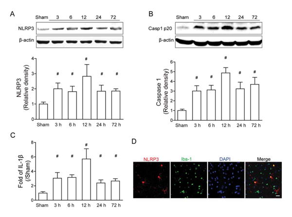
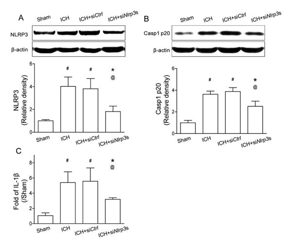
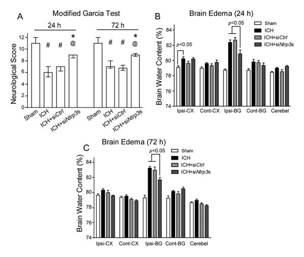
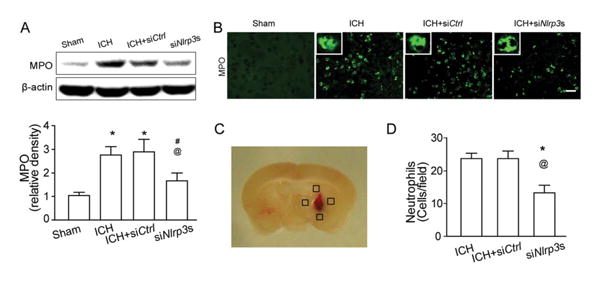
Objective: The NLRP3 (NALP3, cryopyrin) inflammasome, a key component of the innate immune system, facilitates caspase-1 and interleukin (IL)-1β processing, which amplifies the inflammatory response. Here, we investigated whether NLRP3 knockdown decreases neutrophil infiltration, reduces brain edema, and improves neurological function in an intracerebral hemorrhage (ICH) mouse model. We also determined whether mitochondrial reactive oxygen species (ROS) governed by mitochondrial permeability transition pores (mPTPs) would trigger NLRP3 inflammasome activation following ICH.

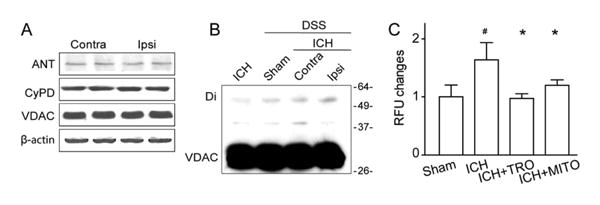
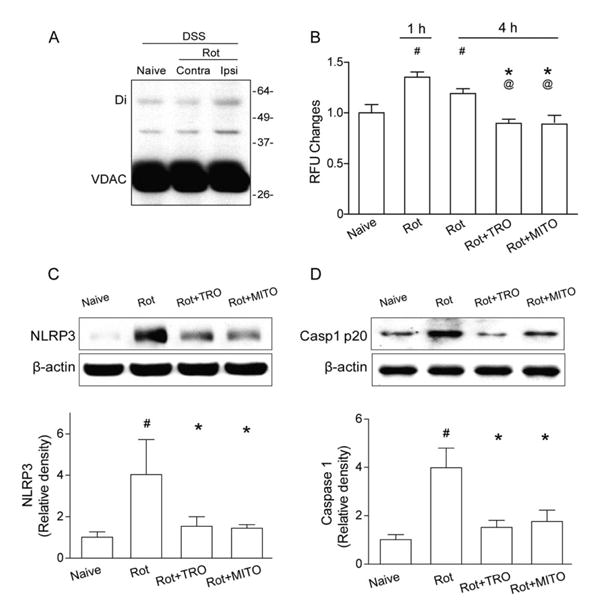
Methods: ICH was induced by injecting autologous arterial blood (30μl) into a mouse brain. NLRP3 small interfering RNAs were administered 24 hours before ICH. A mPTP inhibitor (TRO-19622) or a specific mitochondria ROS scavenger (Mito-TEMPO) was coinjected with the blood. In naive animals, rotenone, which is a respiration chain complex I inhibitor, was applied to induce mitochondrial ROS production, and followed by TRO-19622 or Mito-TEMPO treatment. Neurological deficits, brain edema, enzyme-linked immunosorbent assay, Western blot, in vivo chemical cross-linking, ROS assay, and immunofluorescence were evaluated.

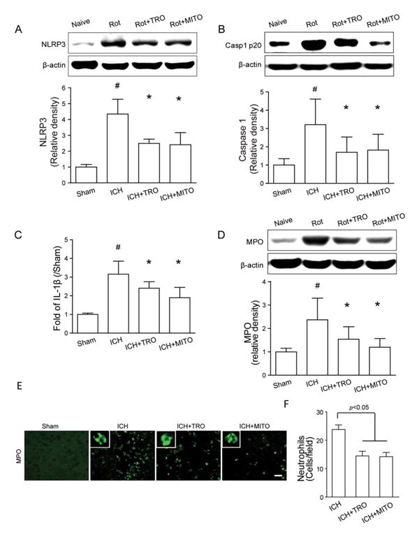
Results: ICH activated the NLRP3 inflammasome. NLRP3 knockdown reduced brain edema and decreased myeloperoxidase (MPO) levels at 24 hours, and improved neurological functions from 24 to 72 hours following ICH. TRO-19622 or Mito-TEMPO reduced ROS, NLRP3 inflammasome components, and MPO levels following ICH. In naive animals, rotenone administration induced mPTP formation, ROS generation, and NLRP3 inflammasome activation, which were then reduced by TRO-19622 or Mito-TEMPO.

Interpretation: The NLRP3 inflammasome amplified the inflammatory response by releasing IL-1β and promoting neutrophil infiltration following ICH. Mitochondria ROS may be a major trigger of NLRP3 inflammasome activation. The results of our study suggest that the inhibition of the NLRP3 inflammasome may effectively reduce the inflammatory response following ICH.ANN NEUROL 2014;75:209-219.


Conflict of interest statement

Potential Conflicts of Interest: Nothing to report.

Figures

Figure 1
Figure 1
Expression profile of the NLRP3 inflammasome components after autologous arterial blood–induced intracerebral hemorrhage (ICH). (A–C) Western blot assay for the expression profiles of NLRP3 (A) and caspase-1 p20 subunit (B), and enzyme-linked immunosorbent assay assay for the expression profiles of interleukin (IL)–1β (C) in the ipsilateral hemisphere in sham and ICH mice at 3, 6, 12, 24, and 72 hours following operation; n=6 mice per group and per time point. Error bars represent mean±standard error of the mean. #p<0.05 vs sham. (D) Representative photographs of immunofluorescence staining for NLRP3 (green) expression in microglia (Iba-1, red) in the perihematomal area 12 hours following ICH. Scale bar=50μm.
Figure 2
Figure 2
NLRP3 small interfering RNA mixture knocked down the NLRP3 inflammasome components at 12 hours following intracerebral hemorrhage (ICH). (A) Western blot assay for the expression of NLRP3 and (B) caspase-1 p20 subunit, and (C) enzyme-linked immunosorbent assay for the expression of interleukin (IL)–1β in the ipsilateral hemisphere in sham, ICH, scrambled siRNA (ICH+siCtrl), and NLRP3 siRNA mixture (ICH+siNlrp3s) groups at 12 hours following operation; n=6 mice per group. Error bars represent mean±standard error of the mean. #p<0.05 vs sham, *p<0.05 vs ICH, @p<0.05 vs ICH+ siCtrl.
Figure 3
Figure 3
NLRP3 knockdown improved neurological functions and reduced brain edema at 24 and 72 hours following intracerebral hemorrhage (ICH). (A) Modified Garcia test at 24 and 72 hours following operation in sham, ICH, scrambled small interfering RNA (siRNA; ICH+siCtrl), and NLRP3 siRNA mixture (ICH+siNlrp3s) groups at 12 hours following operation. (B, C) Brain edema at 24 hours (B) and 72 hours (C) following operation in sham, ICH, scrambled siRNA (ICH+siCtrl), and NLRP3 siRNA mixture (ICH+siNlrp3s) groups at 12 hours following operation. Brain sections (4mm) were divided into 4 parts: ipsilateral basal ganglia (Ipsi-BG), ipsilateral cortex (Ipsi-CX), contralateral basal ganglia (Cont-BG), and contralateral cortex (Cont-CX). Cerebellum (Cerebel) is the internal control; n=6–13 mice per group. Error bars represent median±25th to 75th percen-tiles (A) or mean±standard error of the mean (B, C). #p<0.05 vs sham, *p<0.05 vs ICH, @p<0.01 vs ICH+siCtrl.
Figure 4
Figure 4
NLRP3 knockdown–attenuated neutrophils infiltration at 24 hours following intracerebral hemorrhage (ICH). (A) Western blot assay for the myeloperoxidase (MPO) level in the ipsilateral hemisphere in sham, ICH, scrambled small interfering RNA (siRNA; ICH+siCtrl), and NLRP3 siRNA mixture (ICH+siNlrp3s) groups at 12 hours following operation; n=9 mice per group. Error bars represent mean±standard error of the mean. *p<0.05 vs sham; #p<0.05 vs ICH; @p<0.05 vs ICH+siCtrl. (B) Representative photographs of immunofluorescence staining for MPO-positive cells (green) in the perihematomal area in sham, ICH, scrambled siRNA (ICH+siCtrl), and NLRP3 siRNA mixture (ICH+siNlrp3s) groups 12 hours following ICH. Scale bar=50μm. (C) The schematic diagram shows the 4 areas (black squares) for the MPO-positive cell counting in the perihemato-mal region. (D) Bar graph illustrates the quantification of MPO-positive cells in the perihematomal region in ICH, scrambled siRNA (ICH+siCtrl), and NLRP3 siRNA mixture (ICH+siNlrp3s) groups at 24 hours following operation (12 fields/brain); n=6 mice per group. Error bars represent mean±standard error of the mean. *p<0.05 vs ICH, @p<0.01 vs ICH+siCtrl. [Color figure can be viewed in the online issue, which is available at www.annalsofneurology.org.]
Figure 5
Figure 5
Mitochondrial reactive oxygen species ROS (mROS) production was associated with the formation of mitochondrial permeability transition pore (mPTP) protein assemblies. (A) Western blot assay for the 3 core components of the mPTP (voltage-dependent anion channel [VDAC], adenine nucleotide translocase [ANT], and cyclophilin D [CypD]) in the ipsilateral (Ipsi) versus contralateral (Contra) hemisphere at 12 hours following intracerebral hemorrhage (ICH); n=4 mice per group. (B) Western blots for VDAC in ICH without or with chemical cross-linker (disuccinimidyl suberate [DSS]) at 12 hours following operation. Monomeric VDAC is located at 30kDa. Dimeric (Di) VDAC is detected around 60kDa. (C) Quantification of ROS changes in the perihematomal region in sham, ICH, ICH+TRO-19622 (ICH+TRO), and ICH+Mito-TEMPO (ICH+MITO) groups at 12 hours following operation; n=6 mice per group. Error bars represent mean±standard error of the mean. #p<0.05 vs sham, *p<0.05 vs ICH. RFU=relative fluorescence units.
Figure 6
Figure 6
Mitochondrial reactive oxygen species inhibition reduced NLRP3 inflammasome components expression and neutrophil infiltration following intracerebral hemorrhage (ICH). (A–C) Western blot assay for the expression of NLRP3 (A) and caspase-1 p20 subunit (B), and enzyme-linked immunosorbent assay for the expression of interleukin (IL)–1β (C) in the ipsilateral hemisphere in sham, ICH, ICH+TRO-19622 (ICH+TRO), and ICH+Mito-TEMPO (ICH+MITO) groups at 12 hours following operation; n=6 mice per group. Rot=rotenone injection. Error bars represent mean±standard error of the mean. #p<0.05 vs sham, *p<0.05 vs ICH. (D) Western blot assay for the myeloperoxidase (MPO) level in the ipsilateral hemisphere in sham, ICH, ICH+TRO, and ICH+MITO groups at 12 hours following operation; n=6 mice per group. Error bars represent mean±standard error of the mean. #p<0.05 vs sham, *p<0.05 vs ICH. (E) Representative photographs of immunofluorescence staining for MPO-positive cells (green) in the perihematomal area in sham, ICH, ICH+TRO, and ICH+MITO groups at 12 hours following operation. Scale bar= 50μm. (F) Bar graph illustrating the quantification of MPO-positive cells in the perihematomal region in ICH, ICH+TRO, and ICH+MITO groups at 24 hours following operation (12 fields/brain); n=6 mice per group. Error bars represent mean±standard error of the mean. [Color figure can be viewed in the online issue, which is available at www.annalsofneurology.org.]
Figure 7
Figure 7
Mitochondrial reactive oxygen species (mROS) promotes mitochondrial permeability transition pore formation and NLRP3 inflammasome component expression in naive animals. (A) Western blots for voltage-dependent anion channel (VDAC) in naive and intracerebral hemorrhage (ICH) animals with chemical cross-linker (disuccinimidyl suberate [DSS]) at 1 hour following rotenone injection (Rot). Monomeric VDAC is located at 33kDa. Dimeric (Di) VDAC is detected at 45kDa. Contra=contra-lateral hemisphere; Ipsi = ipsilateral hemisphere. (B) Quantification of mROS alterations in the ipsilateral hemisphere in naive mice, 1 or 4 hours following rotenone injection (Rot), or TRO-19622 (Rot+TRO) or Mito-TEMPO treatment (Rot+MITO) 4 hours following rotenone injection; n=6 mice per group. Error bars represent mean±standard error of the mean. #p<0.05 vs naive, *p<0.05 vs Rot 1 hour; @p<0.05 vs Rot 4 hours. (C, D) Western blot assay for the expression of NLRP3 (C) and caspase-1 p20 subunit (D) in the ipsilateral hemisphere in naive, Rot, ICH+TRO, and ICH+MITO groups at 4 hours following operation; n=6 mice per group. #p<0.05 vs naive, *p<0.05 vs Rot.

Comment in

  • Reactive oxygen species and NLRP3 inflammasome activation.
    Li J, Yan F, Chen G. Li J, et al. Ann Neurol. 2014 Jun;75(6):972. doi: 10.1002/ana.24173. Epub 2014 May 28. Ann Neurol. 2014. PMID: 24798299 No abstract available.
  • Reply: To PMID 24273204.
    Ma Q, Chen S, Hu Q, Feng H, Zhang JH, Tang J. Ma Q, et al. Ann Neurol. 2014 Jun;75(6):972-973. doi: 10.1002/ana.24175. Epub 2014 May 28. Ann Neurol. 2014. PMID: 24805252 Free PMC article. No abstract available.

References

    1. Ribo M, Grotta JC. Latest advances in intracerebral hemorrhage. Curr Neurol Neurosci Rep. 2006;6:17–22. - PubMed
    1. Fisher M, Vasilevko V, Cribbs DH. Mixed cerebrovascular disease and the future of stroke prevention. Transl Stroke Res. 2012;3(suppl 1):39–51. - PMC - PubMed
    1. Qureshi AI, Tuhrim S, Broderick JP, et al. Spontaneous intracerebral hemorrhage. N Engl J Med. 2001;344:1450–1460. - PubMed
    1. Lively S, Schlichter LC. Age-related comparisons of evolution of the inflammatory response after intracerebral hemorrhage in rats. Transl Stroke Res. 2012;3(suppl 1):132–146. - PMC - PubMed
    1. Lok J, Zhao S, Leung W, et al. Neuregulin-1 effects on endothelial and blood-brain-barrier permeability after experimental injury. Transl Stroke Res. 2012;3(suppl 1):S119–S124. - PMC - PubMed

Publication types

MeSH terms