Review
. 2013 Nov 20:4.
doi: 10.3402/dfa.v4i0.21884. eCollection 2013.

Charcot foot in diabetes and an update on imaging

Affiliations
Review

Charcot foot in diabetes and an update on imaging

Fatma Bilge Ergen et al. Diabet Foot Ankle. .

Abstract

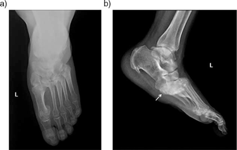
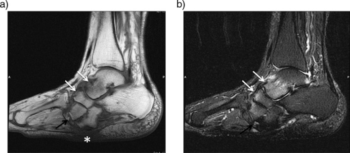
Charcot neuroarthropathy (CN) is a serious complication of diabetes mellitus that can cause major morbidity including limb amputation. Since it was first described in 1883, and attributed to diabetes mellitus in 1936, the diagnosis of CN has been very challenging even for the experienced practitioners. Imaging plays a central role in the early and accurate diagnosis of CN, and in distinction of CN from osteomyelitis. Conventional radiography, computed tomography, nuclear medicine scintigraphy, magnetic resonance imaging, and positron emission tomography are the imaging techniques currently in use for the evaluation of CN but modalities other than magnetic resonance imaging appeared to be complementary. This study focuses on imaging findings of acute and chronic neuropathic osteoarthropathy in diabetes and discrimination of infected vs. non-infected neuropathic osteoarthropathy.

Keywords: Charcot foot; complications; diabetes mellitus; diabetic foot; diagnostic imaging.


Figures

Fig. 1
Fig. 1
Illustration of Sanders and Frykberg's classification of CN. Pattern I: phalanges, interphalangeal and the metatarsophalangeal joints; pattern II: the tarsometatarsal joints; pattern III: the cuneonavicular, talonavicular, and calcaneocuboid articulations; pattern IV: the talocrural joint; pattern V: the posterior calcaneal involvement.
Fig. 2
Fig. 2
Acute neuropathic arthropathy of the foot of 53-year-old woman with diabetes. Sagittal T1 (a) and T2-weighted fat saturated images (b) reveal diffuse bone marrow edema around the Lisfranc joint and calcaneus (arrows). There is also a subcutaneous soft tissue edema especially in the dorsum of the foot. Contrast enhanced sagittal T1-weighted image (c) shows increased enhancement in bone marrow and periarticular tissue (arrows). No evidence of fluid collection or sinus tract noted.
Fig. 3
Fig. 3
Acute neuroarthropathy in an 80-year-old man with long-standing diabetes. Sagittal (a), and coronal (b) T2-weighted fat-saturated images demonstrated massive soft tissue edema in subcutaneous and periarticular region and periarticular bone marrow edema around the Lisfranc joints (black arrow) and posterior subtalar joint. Also note tiny cysts (solid white arrows) along the posterior subtalar joint. Sagittal (c) and coronal (d) post-contrast T1-weighted fat-saturated images demonstrated strong enhancement in the periartricular bone marrow (black arrows) and in the soft tissue planes (white arrows). Late phase bone scintigraphy (e) reveals an increased uptake along the affected bone and joints (arrow).
Fig. 4
Fig. 4
Bone scan of a 53-year-old woman with a history of diabetes presented with swollen right with no skin ulcer. Three-phase (early, blood pool and delayed static phases) hydroxymethane diphosphonate (99mTc-HDP) bone scan demonstrated an increased uptake in all three phases, which is suggestive for diabetic neuroarthropathy (arrows).
Fig. 5
Fig. 5
Anteroposterior (a) and lateral (b) plain radiographs of the left foot in a diabetic patient with chronic neuropathic arthropathy in the midfoot affecting the Lisfranc joint. Note plantar subluxation of the hindfoot (arrow) with typical ‘rocker-bottom’ deformity and dorsal subluxation of metatarsal bases.
Fig. 6
Fig. 6
Midfoot reconstruction in a 60-year-old patient with unstable neuropathic osteoarthropathy (a–c). Anteroposterior (a) and lateral (b) plain radiographs demonstrated neuropathic changes in midfoot region and complex realignment and fusion (a). Joint destruction osseous fragmentation and subchondral cyst formation (arrow) better delineated compared to PR in transverse CT image (c).
Fig. 7
Fig. 7
Anteroposterior (a) and lateral (b) plain radiographs of the foot in a 57-year-old diabetic patient with longstanding neuropathic arthropathy show disorganization and fragments (white arrows) along Lisfranc and Chopart joints. Sagittal T1-weighted (c) and T2-weighted fat-saturated (d) images demonstrate periarticular bone marrow edema (white arrows), periarticular soft tissue collections (black arrow) in Chopart joint. Long-axis post-contrast T1-weighted image (e) reveals periarticular rim enhancing cyst formations (white arrows).
Fig. 8
Fig. 8
Subacute neuroarthropathy of a 60-year-old woman. Sagittal T1-weighted (a) and T2-weighted fat-suppressed image (b) demonstrates rocker-bottom deformity. There is mild bone marrow edema in talonavicular and tarsometatarsal joints (white arrows) and bony fragments adjacent to lateral cuneiform (black arrows). Note the plantar skin callus beneath the cuboid due to altered biomechanics (asterisk).
Fig. 9
Fig. 9
A 57- year-old diabetic patient with longstanding neuropathic arthropathy. Plain radiograph shows hindfoot deformity and calcaneal fragmentation due to Charcot disease (a). T1-weighted sagittal pre (b) and post-contrast (c) and T2-weighted fat-suppressed (d) images reveal skin ulcer and associated a wide sinus tract formation (arrow) containing free air (asteriks), extending to bony cortex. Note the replacement of soft tissue fat (solid white arrow); subcutaneous and periarticular enhancement and osseous contrast enhancement that is consistent with infected neuroarthropathy.

References

    1. Charcot J. Sur quelques arthropathies qui paraissent dependre d'une lesion du cerveau ou de la moelle epiniere. Arch Des Phys Norm Pathol. 1868;1:161.
    1. Gouveri E, Papanas N. Charcot osteoarthropathy in diabetes: a brief review with an emphasis on clinical practice. World J Diabetes. 2011;2:59–65. - PMC - PubMed
    1. Malhotra S, Bello E, Kominsky S. Diabetic foot ulcerations: biomechanics, charcot foot, and total contact cast. Semin Vasc Surg. 2012;25:66–9. - PubMed
    1. Mumoli N, Camaiti A. Charcot foot. CMAJ. 2012;184:1392. - PMC - PubMed
    1. Rogers LC, Frykberg RG, Armstrong DG, Boulton AJ, Edmonds M, Van GH, et al. The Charcot foot in diabetes. Diabetes Care. 2011;34:2123–9. - PMC - PubMed

LinkOut - more resources