

Cerebral magnetic resonance elastography in supranuclear palsy and idiopathic Parkinson's disease
- PMID: 24273721
- PMCID: PMC3814959
- DOI: 10.1016/j.nicl.2013.09.006
Cerebral magnetic resonance elastography in supranuclear palsy and idiopathic Parkinson's disease
Abstract
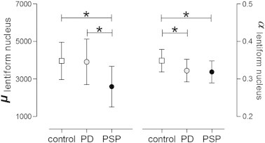
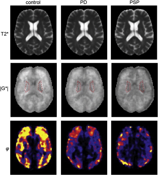
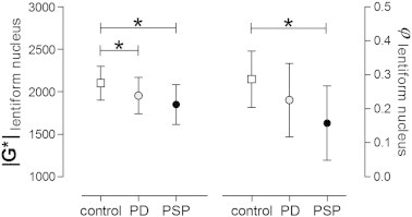
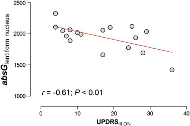
Detection and discrimination of neurodegenerative Parkinson syndromes are challenging clinical tasks and the use of standard T1- and T2-weighted cerebral magnetic resonance (MR) imaging is limited to exclude symptomatic Parkinsonism. We used a quantitative structural MR-based technique, MR-elastography (MRE), to assess viscoelastic properties of the brain, providing insights into altered tissue architecture in neurodegenerative diseases on a macroscopic level. We measured single-slice multifrequency MRE (MMRE) and three-dimensional MRE (3DMRE) in two neurodegenerative disorders with overlapping clinical presentation but different neuropathology - progressive supranuclear palsy (PSP: N = 16) and idiopathic Parkinson's disease (PD: N = 18) as well as in controls (N = 18). In PSP, both MMRE (Δμ = - 28.8%, Δα = - 4.9%) and 3DMRE (Δ|G*|: - 10.6%, Δφ: - 34.6%) were significantly reduced compared to controls, with a pronounced reduction within the lentiform nucleus (Δμ = - 34.6%, Δα = - 8.1%; Δ|G*|: - 7.8%, Δφ: - 44.8%). MRE in PD showed a comparable pattern, but overall reduction in brain elasticity was less severe reaching significance only in the lentiform nucleus (Δμ n.s., Δα = - 7.4%; Δ|G*|: - 6.9%, Δφ: n.s.). Beyond that, patients showed a close negative correlation between MRE constants and clinical severity. Our data indicate that brain viscoelasticity in PSP and PD is differently affected by the underlying neurodegeneration; whereas in PSP all MRE constants are reduced and changes in brain softness (reduced μ and |G*|) predominate those of viscosity (α and φ) in PD.
Keywords: Elasticity; MR-elastography; MRE; Parkinson disease; Progressive supranuclear palsy; Viscosity.
Figures
References
-
- Armstrong R.A., Cairns N.J. Spatial patterns of the tau pathology in progressive supranuclear palsy. Neurol. Sci. 2013;34(3):337–344. - PubMed
-
- Asbach P., Klatt D., Schlosser B., Biermer M., Muche M., Rieger A., Loddenkemper C., Somasundaram R., Berg T., Hamm B., Braun J., Sack I. Viscoelasticity-based staging of hepatic fibrosis with multifrequency MR elastography. Radiology. 2010;257:80–86. - PubMed
-
- Baghani A., Salcudean S., Honarvar M., Sahebjavaher R.S., Rohling R., Sinkus R. Travelling wave expansion: a model fitting approach to the inverse problem of elasticity reconstruction. IEEE Trans. Med. Imaging. 2011;30:1555–1565. - PubMed
LinkOut - more resources
Full Text Sources
Other Literature Sources
Research Materials
Miscellaneous
