. 2013 Nov 25:11:117.
doi: 10.1186/1741-7007-11-117.

Lung mesenchymal expression of Sox9 plays a critical role in tracheal development

Affiliations

Lung mesenchymal expression of Sox9 plays a critical role in tracheal development

Gianluca Turcatel et al. BMC Biol. .

Abstract

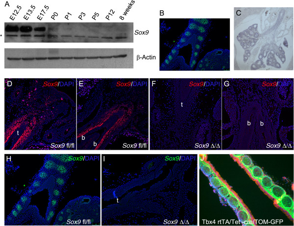
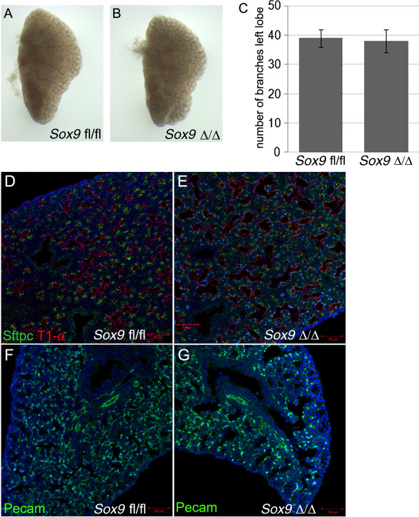
Background: Embryonic lung development is instructed by crosstalk between mesenchyme and epithelia, which results in activation of transcriptional factors, such as Sox9, in a temporospatial manner. Sox9 is expressed in both distal lung epithelium and proximal lung mesenchyme. Here, we investigated the effect of lung mesenchyme-specific inducible deletion of Sox9 during murine lung development.

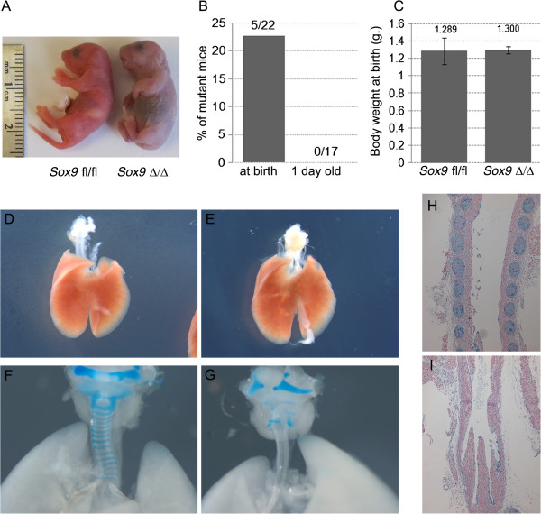
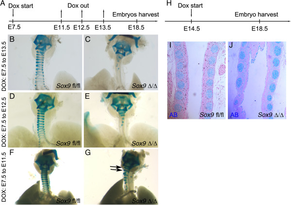
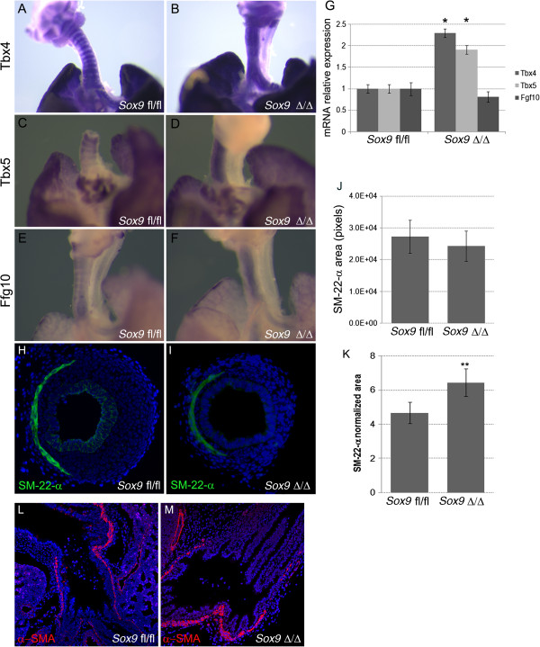
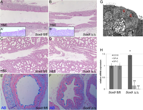
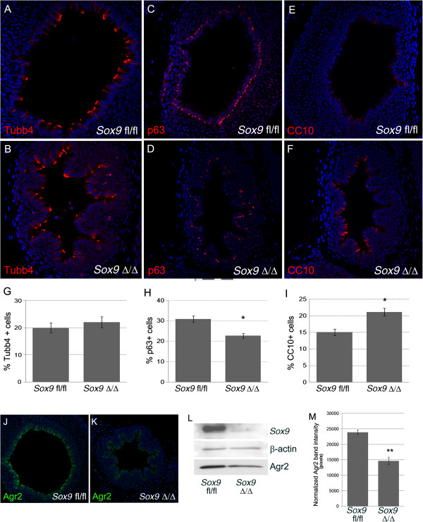
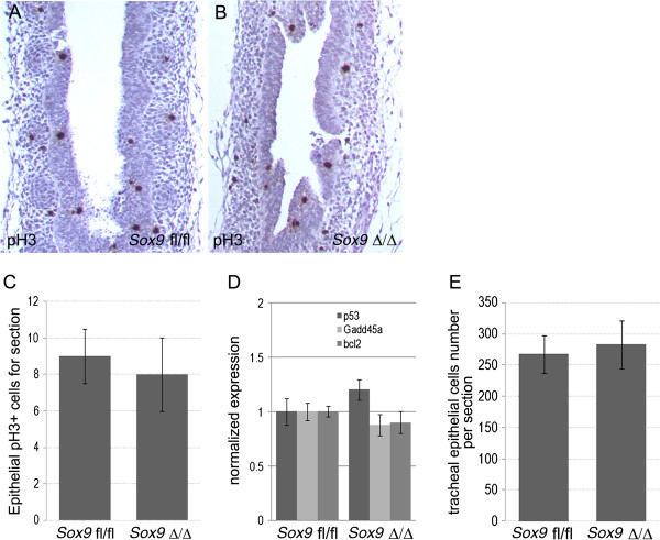
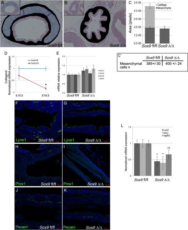
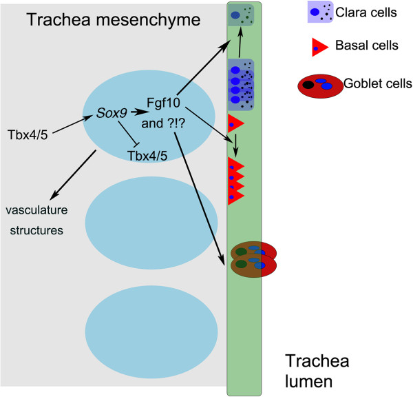
Results: Transgenic mice lacking Sox9 expression were unable to breathe and died at birth, with noticeable tracheal defects. Cartilage rings were missing, and the tracheal lumen was collapsed in the mutant trachea. In situ hybridization showed an altered expression pattern of Tbx4, Tbx5 and Fgf10 genes and marked reduction of Collagen2 expression in the tracheal mesenchyme. The tracheal phenotype was increasingly severe, with longer duration of deletion. Lymphatic vasculature was underdeveloped in the mutant trachea: Prox1, Lyve1, and Vegfr3 were decreased after Sox9 knockout. We also found that compared with normal tracheal epithelium, the mutant tracheal epithelium had an altered morphology with fewer P63-positive cells and more CC10-positive cells, fewer goblet cells, and downregulation of surfactant proteins A and C.

Conclusion: The appropriate temporospatial expression of Sox9 in lung mesenchyme is necessary for appropriate tracheal cartilage formation, lymphatic vasculature system development, and epithelial differentiation. We uncovered a novel mechanism of lung epithelium differentiation: tracheal cartilage rings instruct the tracheal epithelium to differentiate properly during embryonic development. Thus, besides having a mechanical function, tracheal cartilage also appears to be a local signaling structure in the embryonic lung.


Figures

Figure 1
Figure 1
Expression of Sox9 during lung development. (A) Western blot analysis of Sox9 protein expression during lung development at different stages (*non-specific bands). (B)Sox9 protein expression at E15.5 stage: immunofluorescence localization of Sox9 showed expression of Sox9 by tracheal immature cartilage and distal lung epithelium. (C) Expression pattern of endogenous Tbx4 gene in E15.5 lung: Tbx4 is expressed by tracheal mesenchyme and by most, but not all of the distal lung mesenchyme. (D-I)Sox9 immunostaining, showing effective deletion of mesenchyme Sox-9 expression in the mutant lungs at embryonic day (D-G) E12.5 and (H, I) E15.5 stage. (J) Cell tracing using Tbx4-rtTA line. Tbx4-rtTA/Tet-on-cre males were bred with mT/mG females. E7.5 staged pregnant females were fed with doxocycline, and E18.5 embryos were collected. Lung epithelial cells expressed red fluorescent protein, while the mesenchyme cells are green because they are derived from Tbx4-expressing cells (t, trachea; b, bronchus).
Figure 2
Figure 2
Lung phenotype after mesenchymal Sox9 knockout. (A) Pups were collected at birth. Mutant pups died less than an hour after birth, and showed gasping, retractions, and cyanosis. (B) Mutant pups did not survive post-natally. (C) Despite the respiratory mortality phenotype, no change in weight at birth was observed between the wild-type pups and the mutant pups. (D-I) Samples of lung at embryonic day (E) 18.5 were collected and analyzed. (E)Sox9Δ/Δ did not show obvious altered lung branching compared with (D) the control. (F, G) Alcian blue staining of (F) E18.5 wild-type and (G) mutant lungs: normal tracheal rings were observed in the control normal lung, Sox9 knockout trachea revealed no tracheal rings. (H, I) Transverse section of E18.5 trachea was stained with Alcian blue. (H) Wild-type and (I) mutant mice.
Figure 3
Figure 3
Mesenchymal Sox9 deletion did not alter lung branching. (A, C) Left lobes of (A) wild-type and (B) mutant lung at embryonic day (E) 15.5. Terminal branches were counted, and no difference in branching morphogenesis was identified. (D-G) Immunostaining for Sftpc, T1-αc, and Pecam in E18.5 lungs confirmed that distal lung epithelium and mesenchyme differentiation were not altered by Sox9 deletion in the lung mesenchyme.
Figure 4
Figure 4
Doxycycline induction time length affects the severity of trachea phenotype in the Sox9 knockout lungs. (A) Time-plugged female mice were fed with doxycycline for different time periods, and lungs were collected at embryonic day (E) 18.5. (B-G) Doxycycline induction from (B, C) E7.5 to E13.5, (D, E) E7.5 to E12.5, and (F, G) E7.5 to E11.5. (C, E, G) The severity of the phenotype in the transgenic embryos was inversely correlated with the time length of induction. (B, D, F) No changes were observed in the wild-type lung tracheas. (H-J) E14.5 staged pregnant females were fed with doxycycline. Embryos were harvested at E18.5, and the tissues fixed and stained with Alcian blue to show the cartilage rings. (D) Normal cartilage rings developed in the trachea of the Sox9Δ/Δ embryos.
Figure 5
Figure 5
Tbx4, Tbx5, and Fgf10 in situ hybridization results.In situ hybridization for (A, B)Tbx4 and (C, D)Tbx5 was performed on lung at embryonic day (E) 15.5 to identify changes in expression patterns. Tbx4 andTbx5 were expressed by the mesenchyme surrounding the tracheal rings in the normal control embryonic lung tracheas (A, C), whereas Sox9 knockout lungs displayed diffuse expression of (B)Tbx4 and (D)Tbx5 along all the tracheal mesenchyme. (E, F) The Fgf10 expression pattern was lost in the Sox9Δ/Δ trachea. (G) Real time RT-qPCR PCR was used to determine whether expression of Tbx4 and Tbx5 mRNA was increased in the mutant mouse trachea versus wild-type mouse trachea (*P < 0.05). Fgf10 expression was not significantly different between by Sox9Δ/Δ vs. the Sox9fl/fl tracheas. (H-K) Trachealis smooth muscle cells did not change after Sox9 knockout. Staining for Sm-22-α was used to identify the trachealis smooth muscle cells on transverse sections of normal and transgenic mouse tracheas. (J, K) Even though the absolute area of trachealis smooth muscle cells did not change, the Sm-22-α relative area was significantly higher in the Sox9Δ/Δ(J) compared with (K) the Sox9fl/fl trachea, because of the smaller transversal area of the mutant trachea. **P = 0.015. Values are mean ± SD, n = 5. (L, M). Staining for α-smooth muscle actin (α-SMA) was used to highlight smooth muscle cells. Sox9 deletion did not affect lung smooth muscle cell proliferation or differentiation.
Figure 6
Figure 6
Lung tracheal epithelium is altered in Sox9 knockout mice. (A-B) Hematoxylin and eosin (H&E) staining of embryonic day (E) 18.5 tracheal sections, showing altered morphology of epithelial cells in (B) the mutant trachea compared with (A) the wild-type (A′ and B′ are high magnification inserts of A and B respectively). (C, D) Intralobar sections of main bronchi stained with H&E. No obvious differences are seen between (C) the wild-type and (D) mutant intralobar bronchia. (E, F) Transverse sections of (E) wild-type and (F) mutant mouse tracheas stained with Alcian blue. Mutant tracheas are collapsed because of the lack of structural support normally given by the cartilage rings. (G) Electron microscopy of epithelial cells lining the mutant tracheal revealed the presence of Clara cells. (H) Real time RT-qPCR analysis at E18.5 altered expression of Sftpc, Sftpa, and CC-10 genes. *P < 0.05, **P < 0.01. Values are mean ± SD.
Figure 7
Figure 7
Basal cell numbers are decreased and Clara cell numbers are increased in the mutant mouse trachea. (A-B) Ciliated cell number was not altered in Sox9 knockout mouse tracheas. Staining for β-tubulin-IV on transverse section of wild-type and mutant mouse trachea was used to determine change in ciliated cell numbers. (G) The number of ciliated cells did not change in the mutant mouse trachea versus the wild-type mouse trachea. (C, D) Staining for P63 was used to confirm that the number of basal cells is decreased in the mutant mouse trachea. (E, F) CC10 marker staining was used to highlight the increase in Clara cell number in Sox9 mutant mouse trachea. (G-I) Cell count statistics. *P <0.03. Values are mean ± SD, n = 4. (J-M)Sox9Δ/Δ tracheas had a reduced number of goblet cells. (J, K) Immuno-fluorescence staining and (L) western blot for Agr2 protein were used to determine change in goblet cells after Sox9 knockout. Sox9 knockout resulted in reduced production of Agr2 by the tracheal epithelium, suggesting reduced numbers of goblet cells in the Sox9Δ/Δ trachea. (M) Intensity analysis of Western blot bands. **P < 0.05. Values are mean ± SD.
Figure 8
Figure 8
Tracheal epithelial differentiation and apoptosis was not altered by lack of Sox9 gene. (A, B) Phospho-histone-3 (pH3) staining on longitudinal sections of (A) wild-type and (B) mutant tracheas at embryonic day (E) 15.5 did not reveal any change in epithelial cell proliferation. (C) Number of pH3-positive cells per section was similar between wild-type and mutant mouse trachea. (D) Real-time PCR for the apoptotic markers p53, Bcl2, and Gadd45a on total RNA extracted from E15.5 wild-type and mutant tracheas indicated no change in apoptosis. (E) Tracheal epithelial cells were counted on transverse sections of Sox9fl/fl and Sox9Δ/Δ E18.5 tracheas, revealing no change in cell number between the two groups. Values are mean ± SD.
Figure 9
Figure 9
Lymphatic vascular system was affected by Sox9 deletion. (A-C) Tracheal mesenchyme area quantification analysis did not reveal changes between the wild-type and mutant tracheas (tracheal mesenchyme was stained in black). (C′) The number of mesenchymal cells per section was similar between wild-type and mutant trachea. (D, E) Real time RT-qPCR for Collagen2, Gli1, and Shh on mRNA from wild-type and mutant tracheas at embryonic day (E) 15.5. Col2 expression was decreased in the Sox9Δ/Δ trachea starting at E15.5 stage. No statistically significant differences in expression of Shh, Gli1, Gli2, or Gli3 was observed between Sox9fl/fl and Sox9Δ/Δ. (F-L) The lymphatic vascular system was underdeveloped in the mutant trachea. Immunostaining of the lymphatic markers (F, G) Lyve1, (H, I) Prox1, and (J, K) Pecam revealed that Sox9 deletion resulted in impaired lymphatic vessel formation. (L) Real time RT-qPCR for Prox, Lyve1, and Vegfr3 confirmed that the lymphatic vascular system was not properly developed in the Sox9 knockout tracheas. *P < 0.05, **P = 0.08.
Figure 10
Figure 10
Concept diagram of Sox9 function in lung development.Sox9 acts downstream of Tbx4/5[6]. Sox9 feeds back negatively on Tbx4 and Tbx5 in the tracheal mesenchyme. Cartilage ring formation requires functional Sox9 expression in the tracheal mesenchyme. Lack of cartilage rings induce altered tracheal epithelium differentiation (increased Clara cells, and decreased basal and goblet cells) and defective development of tracheal vasculature structurest.

References

    1. Whitsett JA, Wert SE, Trapnell BC. Genetic disorders influencing lung formation and function at birth. Hum Mol Genet. 2004;13:207–215. doi: 10.1093/hmg/ddh252. - DOI - PubMed
    1. Goo HW. Free-breathing cine CT for the diagnosis of tracheomalacia in young children. Pediatr Radiol. 2013;42:922–928. - PubMed
    1. Kandaswamy C. et al. Severe tracheomalacia in the ICU: identification of diagnostic criteria and risk factor analysis from a case control study. Respir Care. 2013;58:340–347. - PubMed
    1. Goldring MB, Tsuchimochi K, Ijiri K. The control of chondrogenesis. J Cell Biochem. 2006;97:33–44. doi: 10.1002/jcb.20652. - DOI - PubMed
    1. Sala FG. et al. FGF10 controls the patterning of the tracheal cartilage rings via Shh. Development. 2011;138:273–282. doi: 10.1242/dev.051680. - DOI - PMC - PubMed

Publication types

MeSH terms

LinkOut - more resources