Case Reports
. 2013 Dec;19(12):2008-11.
doi: 10.3201/eid1912.130284.

Cerebellar cysticercosis caused by larval Taenia crassiceps tapeworm in immunocompetent woman, Germany

Case Reports

Cerebellar cysticercosis caused by larval Taenia crassiceps tapeworm in immunocompetent woman, Germany

Vasileios Ntoukas et al. Emerg Infect Dis. 2013 Dec.

Abstract

Human cysticercosis caused by Taenia crassiceps tapeworm larvae involves the muscles and subcutis mostly in immunocompromised patients and the eye in immunocompetent persons. We report a successfully treated cerebellar infection in an immunocompetent woman. We developed serologic tests, and the parasite was identified by histologic examination and 12s rDNA PCR and sequencing.

Keywords: Germany; PCR; Taenia crassiceps; central nervous system; cerebellar infection; cysticercosis; immunocompetent; molecular identification; parasites; tapeworm; zoonosis.


Figures

Figure 1
Figure 1
Magnetic resonance (MR) and computed tomographic images of the brain of a 51-year-old woman infected with Taenia crassiceps tapeworm larvae, Germany. A) Transverse view, T1-weighted MR image. The 30 × 30 mm parasitic lesion with perifocal edema is located in the right hemisphere of the cerebellum and caused ataxia, headache, and nausea. The fourth ventricle is compressed. B) Coronal view, T2-weighted MR image. The cyst-like appearance of the parasitic tissue is clearly visible. This lesion can be misinterpreted as cerebral echinococcosis, racemose cysticercosis caused by a Taenia solium tapeworm, or coenurosis. C) Sagittal view, MR image with contrast enhancing agent. D) Transverse view, computed tomographic image after surgery. No residual parasitic masses, only the parenchymal defect in the cerebellum after resection of T. crassiceps tapeworm larvae, are visible.
Figure 2
Figure 2
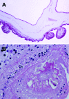
Histologic section through Taenia crassiceps tapeworm larvae removed from the cerebellum of a 51-year-old woman, Germany. A) Section through parasite body showing multiple connected bladders (asexual buddings) at the caudal end. Original magnification ×20. B) Transverse section through the parasite’s protoscolex showing numerous hooklets, similar to T. solium tapeworm larvae. Original magnification ×40. Like the Taenia solium tapeworm that causes cysticercosis, and in contrast to different tapeworms that cause coenurosis (T. [Multiceps] multiceps, T. [Multiceps] serialis) or echinococcosis, the T. crassiceps tapeworm has only 1 invaginated protoscolex, but it is on a very long neck (Cysticercus longicollis). The hooklets of T. crassiceps tapeworms are larger than those of T. solium tapeworms and have a larger blade length than handle length. The small hooklets measure 123 µm; the large hooklets measure 167 µm.

References

    1. Heldwein K, Biedermann HG, Hamperl WD, Bretzel G, Loscher T, Laregina D, et al. Subcutaneous Taenia crassiceps infection in a patient with non-Hodgkin's lymphoma. Am J Trop Med Hyg. 2006;75:108–11 . - PubMed
    1. Bružinskaitė-Schmidhalter R, Sarkunas M, Malakauskas A, Mathis A, Torgerson PR, Deplazes P. Helminths of red foxes (Vulpes vulpes) and raccoon dogs (Nyctereutes procyonoides) in Lithuania. Parasitology. 2012;139:120–7. 10.1017/S0031182011001715 - DOI - PubMed
    1. Saeed I, Maddox-Hyttel C, Monrad J, Kapel CM. Helminths of red foxes (Vulpes vulpes) in Denmark. Vet Parasitol. 2006;139:168–79. 10.1016/j.vetpar.2006.02.015 - DOI - PubMed
    1. Goesseringer N, Lindenblatt N, Mihic-Probst D, Grimm F, Giovanoli P. Taenia crassiceps upper limb fasciitis in a patient with untreated acquired immunodeficiency syndrome and chronic hepatitis C infection—the role of surgical debridement. J Plast Reconstr Aesthet Surg. 2011;64:e174–6 . 10.1016/j.bjps.2011.02.011 - DOI - PubMed
    1. François A, Favennec L, Cambon-Michot C, Gueit I, Biga N, Tron F, et al. Taenia crassiceps invasive cysticercosis: a new human pathogen in acquired immunodeficiency syndrome? Am J Surg Pathol. 1998;22:488–92. 10.1097/00000478-199804000-00015 - DOI - PubMed

Publication types

LinkOut - more resources