Review
. 2013:69:209-72.
doi: 10.1016/B978-0-12-408093-5.00006-X.

Bacterial cell-envelope glycoconjugates

Affiliations
Review

Bacterial cell-envelope glycoconjugates

Paul Messner et al. Adv Carbohydr Chem Biochem. 2013.

Abstract

Prokaryotic glycosylation fulfills an important role in maintaining and protecting the structural integrity and function of the bacterial cell wall, as well as serving as a flexible adaption mechanism to evade environmental and host-induced pressure. The scope of bacterial and archaeal protein glycosylation has considerably expanded over the past decade(s), with numerous examples covering the glycosylation of flagella, pili, glycosylated enzymes, as well as surface-layer proteins. This article addresses structure, analysis, function, genetic basis, biosynthesis, and biomedical and biotechnological applications of cell-envelope glycoconjugates, S-layer glycoprotein glycans, and "nonclassical" secondary-cell wall polysaccharides. The latter group of polymers mediates the important attachment and regular orientation of the S-layer to the cell wall. The structures of these glycopolymers reveal an enormous diversity, resembling the structural variability of bacterial lipopolysaccharides and capsular polysaccharides. While most examples are presented for Gram-positive bacteria, the S-layer glycan of the Gram-negative pathogen Tannerella forsythia is also discussed. In addition, archaeal S-layer glycoproteins are briefly summarized.

Keywords: Archaea; Bacteria; Glycoprotein; Secondary cell-wall polymers; Surface layer.


Figures

Fig. 1
Fig. 1
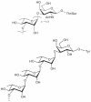
Scheme of cell envelopes of prokaryotic organisms, showing representative cellular glycan structures
Fig. 1
Fig. 1
Scheme of cell envelopes of prokaryotic organisms, showing representative cellular glycan structures
Fig. 2
Fig. 2
Electron micrograph of a freeze-substituted and cross-sectioned cell of Ane. thermoaerophilus DSM 10155. With permission from Springer Science + Business Media, compare with Ref. , Fig. 1b.
Fig. 3
Fig. 3
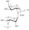
Rhamnosyl core oligosaccharides.-
Fig. 4
Fig. 4
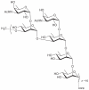
Core structure of P. alvei CCM 2051 S-layer glycan.
Fig. 5
Fig. 5
Core structure of Ane. thermoaerophilus L420-91T.
Fig. 6
Fig. 6
S-Layer glycan of Lb. buchneri.
Fig. 7
Fig. 7
S-Layer glycan structure of G. stearothermophilus NRS 2004/3a.
Fig. 8
Fig. 8
S-Layer glycan structure of Thb. thermohydrosulfuricus strains L111-69 and L110-69.
Fig. 9
Fig. 9
S-Layer glycan structure of Thm. thermosaccharolyticum strain D120-70.
Fig. 10
Fig. 10
S-Layer glycan structure of Ano. tepidamans GS5-97T.
Fig. 11
Fig. 11
S-Layer glycan structure of Ane. thermoaerophilus DSM 10155.
Fig. 12
Fig. 12
S-Layer glycan structure of Ane. thermoaerophilus strains L420-91T and GS4-97.,
Fig. 13
Fig. 13
S-Layer glycan structure of P. alvei CCM 2051T.
Fig. 14
Fig. 14
S-Layer glycan structure of Thb. thermohydrosufuricus L77-66 (DSM 569).
Fig. 15
Fig. 15
S-Layer glycan structure of Thm. thermosaccharolyticum E207-71.
Fig. 16
Fig. 16
S-Layer glycan structure of Thb. thermohydrosulfuricus S102-70.
Fig. 17
Fig. 17
Schematic structure of the T. forsythia S-layer glycan., Adapted from the open access journal Biomolecules; © 2012 by the authors, licensee MDPI, Basel, Switzerland; http://creativecommons.org/licenses/by/3.0.
Fig. 18
Fig. 18
Schematic putative structure of the B. fragilis glycan.
Fig. 19
Fig. 19
Terminal end of the S-layer glycan of Ano. tepidamans GS5-97T.
Fig. 20
Fig. 20
Structure of a glycopeptide from cytochrome b558/566 of Sl. acidocaldarius.
Fig. 21
Fig. 21
Repeating unit of the SCWP from P. alvei CCM 2051T.
Fig. 22
Fig. 22
Structure of the SCWP from Thm. thermosaccharolyticum strain E207-71.
Fig. 23
Fig. 23
Structure of the SCWP from G. stearothermophilus NRS 2004/3a.,
Fig. 24
Fig. 24
Structure of the repeating unit of the SCWP from Ane. thermoaerophilus DSM 10155.
Fig. 25
Fig. 25
Structure of the repeating unit of the acid-degraded SCWP from G. stearothermophilus PV72/p2. Reprinted from Ref. . Copyright (2008), with permission from Elsevier.
Fig. 26
Fig. 26
Structure of HF-treated B. anthracis SCWP.
Fig. 27
Fig. 27
Structure of the HF-treated SCWP from B. cereus strains G92141 and 03BB87.
Fig. 28
Fig. 28
Structure of the HF-treated SCWP from B. cereus ATCC 10987. Adapted from Ref. . © The American Society for Biochemistry and Molecular Biology.
Fig. 29
Fig. 29
Structure of the HF-treated SCWP from B. cereus ATCC 14579.
Fig. 30
Fig. 30
Structure of the charged SCWP from B. cereus ATCC 14579.
Fig. 31
Fig. 31
Biosynthesis pathways for nucleotide sugars as required for S-layer protein glycosylation.,-

Similar articles

  • Biochemistry of S-layers.
    Messner P, Allmaier G, Schäffer C, Wugeditsch T, Lortal S, König H, Niemetz R, Dorner M. Messner P, et al. FEMS Microbiol Rev. 1997 Jun;20(1-2):25-46. doi: 10.1111/j.1574-6976.1997.tb00303.x. FEMS Microbiol Rev. 1997. PMID: 9276927 Review.
  • Emerging facets of prokaryotic glycosylation.
    Schäffer C, Messner P. Schäffer C, et al. FEMS Microbiol Rev. 2017 Jan;41(1):49-91. doi: 10.1093/femsre/fuw036. Epub 2016 Aug 26. FEMS Microbiol Rev. 2017. PMID: 27566466 Free PMC article. Review.
  • Recent advances in mycobacterial cell wall glycan biosynthesis.
    Tam PH, Lowary TL. Tam PH, et al. Curr Opin Chem Biol. 2009 Dec;13(5-6):618-25. doi: 10.1016/j.cbpa.2009.09.012. Epub 2009 Oct 3. Curr Opin Chem Biol. 2009. PMID: 19805002 Review.
  • Glycobiology of surface layer proteins.
    Schäffer C, Messner P. Schäffer C, et al. Biochimie. 2001 Jul;83(7):591-9. doi: 10.1016/s0300-9084(01)01299-8. Biochimie. 2001. PMID: 11522387 Review.
  • Prokaryotic glycosylation.
    Schäffer C, Graninger M, Messner P. Schäffer C, et al. Proteomics. 2001 Feb;1(2):248-61. doi: 10.1002/1615-9861(200102)1:2<248::AID-PROT248>3.0.CO;2-K. Proteomics. 2001. PMID: 11680871 Review.

Cited by

References

    1. Kobata A. The carbohydrates of glycoproteins. In: Ginsburg V, Robbins PW, editors. Biology of Carbohydrates. Vol. 2. John Wiley & Sons; New York: 1984. pp. 87–161.
    1. Montreuil J. Spatial conformation of glycans and glycoproteins. Biol. Cell. 1984;51:115–131. - PubMed
    1. Kornfeld R, Kornfeld S. Assembly of asparagine-linked oligosaccharides. Annu. Rev. Biochem. 1985;54:631–664. - PubMed
    1. Varki A, Cummings RD, Esko JD, Freeze HH, Stanley P, Bertozzi CR, Hart GW, Etzler ME, editors. Essentials of Glycobiology. 2nd ed. Cold Spring Harbor Laboratory Press; Cold Spring Harbor, NY: 2009. p. 784. - PubMed
    1. Messner P, Sleytr UB. Bacterial surface layer glycoproteins. Glycobiology. 1991;1:545–551. - PubMed

LinkOut - more resources