. 2013 Dec 31;52(52):9456-69.
doi: 10.1021/bi4008885. Epub 2013 Dec 17.

Identification of the GPR55 antagonist binding site using a novel set of high-potency GPR55 selective ligands

Affiliations

Identification of the GPR55 antagonist binding site using a novel set of high-potency GPR55 selective ligands

Evangelia Kotsikorou et al. Biochemistry. .

Abstract

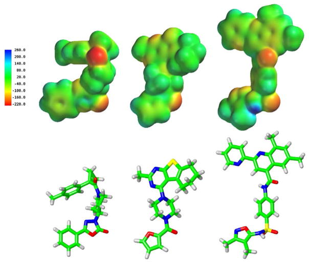
GPR55 is a class A G protein-coupled receptor (GPCR) that has been implicated in inflammatory pain, neuropathic pain, metabolic disorder, bone development, and cancer. Initially deorphanized as a cannabinoid receptor, GPR55 has been shown to be activated by non-cannabinoid ligands such as l-α-lysophosphatidylinositol (LPI). While there is a growing body of evidence of physiological and pathophysiological roles for GPR55, the paucity of specific antagonists has limited its study. In collaboration with the Molecular Libraries Probe Production Centers Network initiative, we identified a series of GPR55 antagonists using a β-arrestin, high-throughput, high-content screen of ~300000 compounds. This screen yielded novel, GPR55 antagonist chemotypes with IC50 values in the range of 0.16-2.72 μM [Heynen-Genel, S., et al. (2010) Screening for Selective Ligands for GPR55: Antagonists (ML191, ML192, ML193) (Bookshelf ID NBK66153; PMID entry 22091481)]. Importantly, many of the GPR55 antagonists were completely selective, with no agonism or antagonism against GPR35, CB1, or CB2 up to 20 μM. Using a model of the GPR55 inactive state, we studied the binding of an antagonist series that emerged from this screen. These studies suggest that GPR55 antagonists possess a head region that occupies a horizontal binding pocket extending into the extracellular loop region, a central ligand portion that fits vertically in the receptor binding pocket and terminates with a pendant aromatic or heterocyclic ring that juts out. Both the region that extends extracellularly and the pendant ring are features associated with antagonism. Taken together, our results provide a set of design rules for the development of second-generation GPR55 selective antagonists.


Figures

Figure 1
Figure 1
GPR55 antagonist structures are shown here. ML191, ML192 and ML193 are the focus of this work. CID16020046 is another GPR55 antagonist recently published (48) by Kargl and co-workers.
Figure 2
Figure 2
An extracellular view of the GPR55 inactive state (R) model is illustrated here. The location of the two extracellular disulfide bridges are indicated.
Figure 3
Figure 3
Antagonist Activity of ML191, ML192 and ML193. Concentration response curves for antagonist activity of ML191, ML192, and ML193 in the β-arrestin trafficking assay using LPI (A) or ML186 (B) as an agonist. Concentration response curves for LPI (C) and ML186 (D).
Figure 4
Figure 4
LPI mediated ERK1/2 phosphorylation is largely inhibited by ML 191–193 compounds in U2OS cells overexpressing GPR55E and βarr2/GFP. Cells were pre-incubated with increasing concentrations of ML compounds 30 min prior to the application of LPI (10 μM). ERK1/2 phosphorylation levels were monitored and normalized to total ERK1/2 levels and IC50 values for each of the compounds was determined. Data are mean ± SEM from three independent experiments performed in duplicate (*P<0.05, **P<0.01, ***P<0.001). LPI mediated ERK1/2 phosphorylation was significantly attenuated following treatment with ML191 (A), ML192(B) and ML193(C). In untransfected U2OS cells, neither LPI nor any of the antagonist compounds induced ERK1/2 phosphorylation, while activation by pervanadate indicated that the MAPK pathway is intact (D).
Figure 5
Figure 5
Molecular electrostatic potential maps of ML191, ML192 and ML193 are shown here in the top row. In the second row, the conformer of each used to calculate the map is shown in tube display.
Figure 6
Figure 6
ML193 (blue) is shown docked in the GPR55 R model. In the view shown here, the top of TMH7 has been omitted for clarity. K2.60(80) (pink) was observed to be a primary interaction site during docking studies. The toggle switch residues M3.36(104)/F6.48(239) are shown in purple. Residues that contributed at least 2.5% to the total interaction energy are shown in orange. See Table S.1 in Supplemental Information for a breakdown of the energy of interaction ofML193 in GPR55 R with specific residues.
Figure 7
Figure 7
ML191 (blue) is shown docked in the GPR55 R model. In the view shown here, the top of TMH7 has been omitted for clarity. K2.60(80) (pink) was observed to be a primary interaction site during docking studies. The toggle switch residues M3.36(104)/F6.48(239) are shown in purple. Residues that contributed at least 2.5% to the total interaction energy are shown in orange. See Table S.2 in Supplemental Information for a breakdown of the energy of interaction of ML191 in GPR55 R broken down by residue.
Figure 8
Figure 8
ML192 docked to GPR55 R. ML192 (blue) is shown docked in the GPR55 R model. In the view shown here, the top of TMH7 has been omitted for clarity. K2.60(80) (pink) was observed to be a primary interaction site during docking studies. The toggle switch residues M3.36(104)/F6.48(239) are shown in purple. Residues that contributed at least 2.5% to the total interaction energy are shown in orange. See Table S.4 in Supplemental Information for a breakdown of the energy of interaction of ML192 in GPR55 R broken down by residue.
Figure 9
Figure 9
MD studies suggested that ML192 was more mobile in the binding pocket such that it could adopt a more extracellular binding region than that of the static ML192/GPR55 R dock. To understand the differences, we extracted a frame from the MD trajectory and energy minimized the system using the same minimization protocol as described earlier. The results are illustrated here. ML192 (blue) is shown docked in the GPR55 R model. In the view shown here, the top of TMH7 has been omitted for clarity. K2.60(80) (pink) was observed to be a primary interaction site during the molecular dynamics simulation. The toggle switch residues M3.36(104)/F6.48(239) are shown in purple. Residues that contributed at least 2.5% to the total interaction energy are shown in orange. See Table S.4 in Supplemental Information for a breakdown of the energy of interaction of ML192 in GPR55 R broken down by residue.
Figure 10
Figure 10
The structures of a GPR55 agonist (CID1792197) (14) and the GPR55 antagonist (ML191) are shown here. The agonist has its most electronegative region (which hydrogen bonds to K2.60) near the broad head region, while this region is found in antagonists near the end of the central portion of the molecule. In addition, agonist structures lack the pendant phenyl ring found in ML191 after the central portion. Instead, agonist structures maintain a thin vertical profile in the binding pocket.

References

    1. Henstridge CM, Balenga NA, Kargl J, Andradas C, Brown AJ, Irving A, Sanchez C, Waldhoer M. Minireview: Recent Developments in the Physiology and Pathology of the Lysophosphatidylinositol-Sensitive Receptor GPR55. Mol Endocrinol. 2011;25:1835–1848. - PMC - PubMed
    1. Staton PC, Hatcher JP, Walker DJ, Morrison AD, Shapland EM, Hughes JP, Chong E, Mander PK, Green PJ, Billinton A, Fulleylove M, Lancaster HC, Smith JC, Bailey LT, Wise A, Brown AJ, Richardson JC, Chessell IP. The putative cannabinoid receptor GPR55 plays a role in mechanical hyperalgesia associated with inflammatory and neuropathic pain. Pain. 2008;139:225–236. - PubMed
    1. Whyte LS, Ryberg E, Sims NA, Ridge SA, Mackie K, Greasley PJ, Ross RA, Rogers MJ. The putative cannabinoid receptor GPR55 affects osteoclast function in vitro and bone mass in vivo. Proc Natl Acad Sci U S A. 2009;106:16511–16516. - PMC - PubMed
    1. Ford LA, Roelofs AJ, Anavi-Goffer S, Mowat L, Simpson DG, Irving AJ, Rogers MJ, Rajnicek AM, Ross RA. A role for L-alpha-lysophosphatidylinositol and GPR55 in the modulation of migration, orientation and polarization of human breast cancer cells. Br J Pharmacol. 2010;160:762–771. - PMC - PubMed
    1. Andradas C, Caffarel MM, Perez-Gomez E, Salazar M, Lorente M, Velasco G, Guzman M, Sanchez C. The orphan G protein-coupled receptor GPR55 promotes cancer cell proliferation via ERK. Oncogene. 2011;30:245–252. - PubMed

Publication types