Review
. 2014:76:333-63.
doi: 10.1146/annurev-physiol-021113-170338. Epub 2013 Nov 21.

Molecular mechanisms for synchronous, asynchronous, and spontaneous neurotransmitter release

Affiliations
Review

Molecular mechanisms for synchronous, asynchronous, and spontaneous neurotransmitter release

Pascal S Kaeser et al. Annu Rev Physiol. 2014.

Abstract

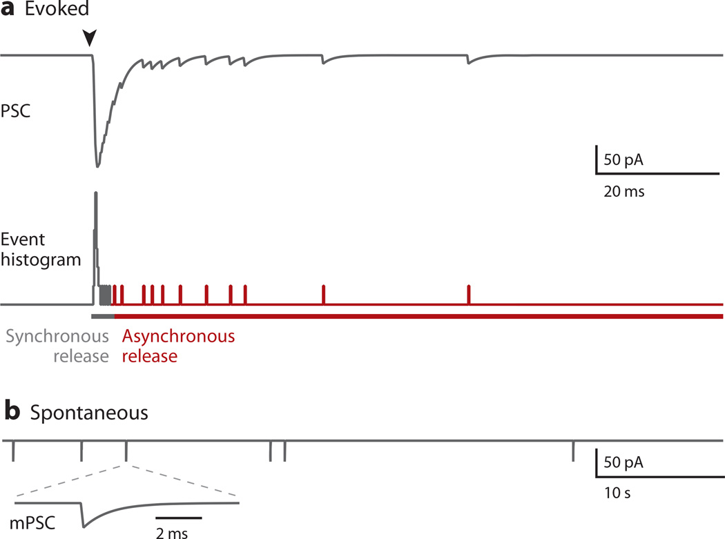
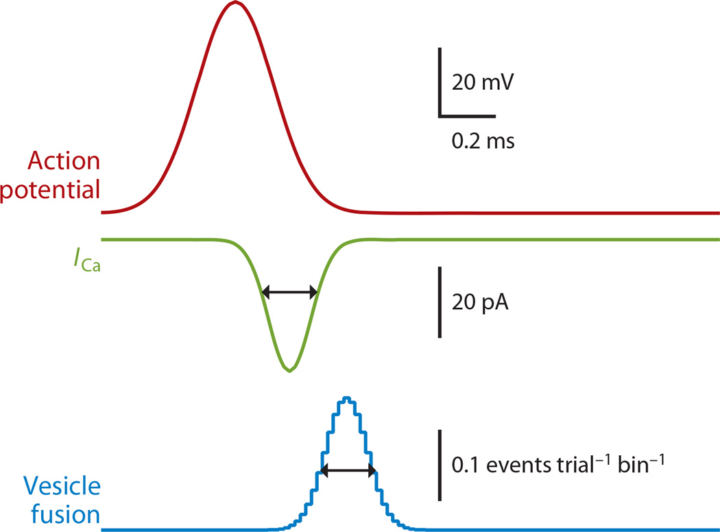
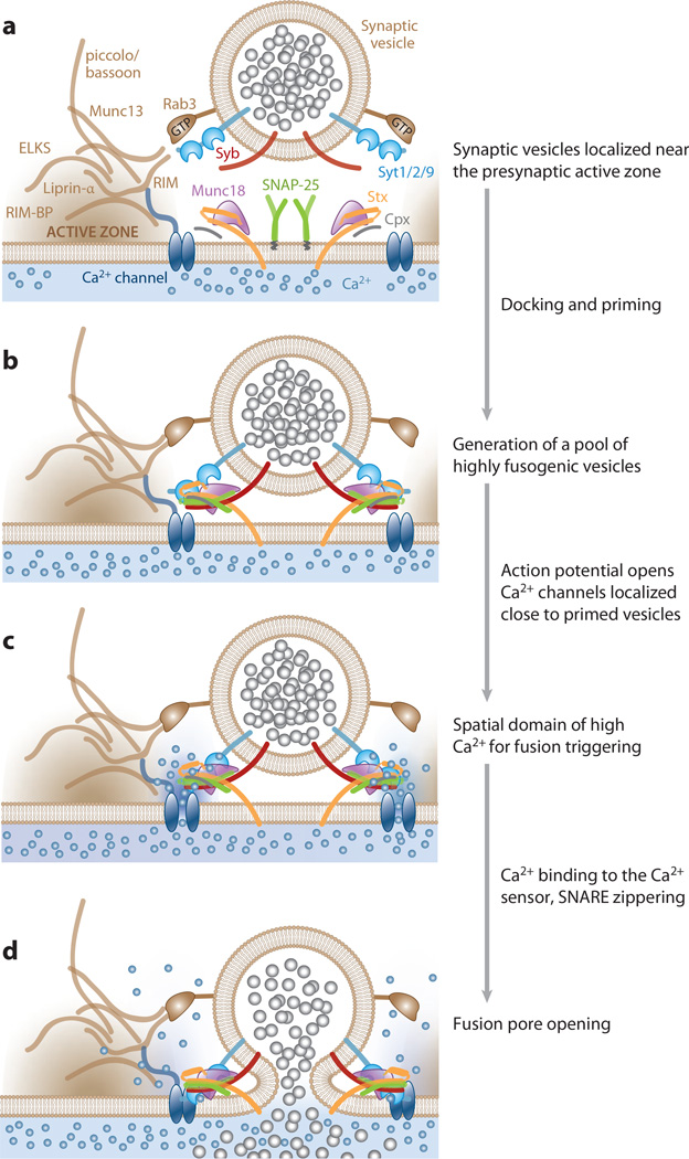
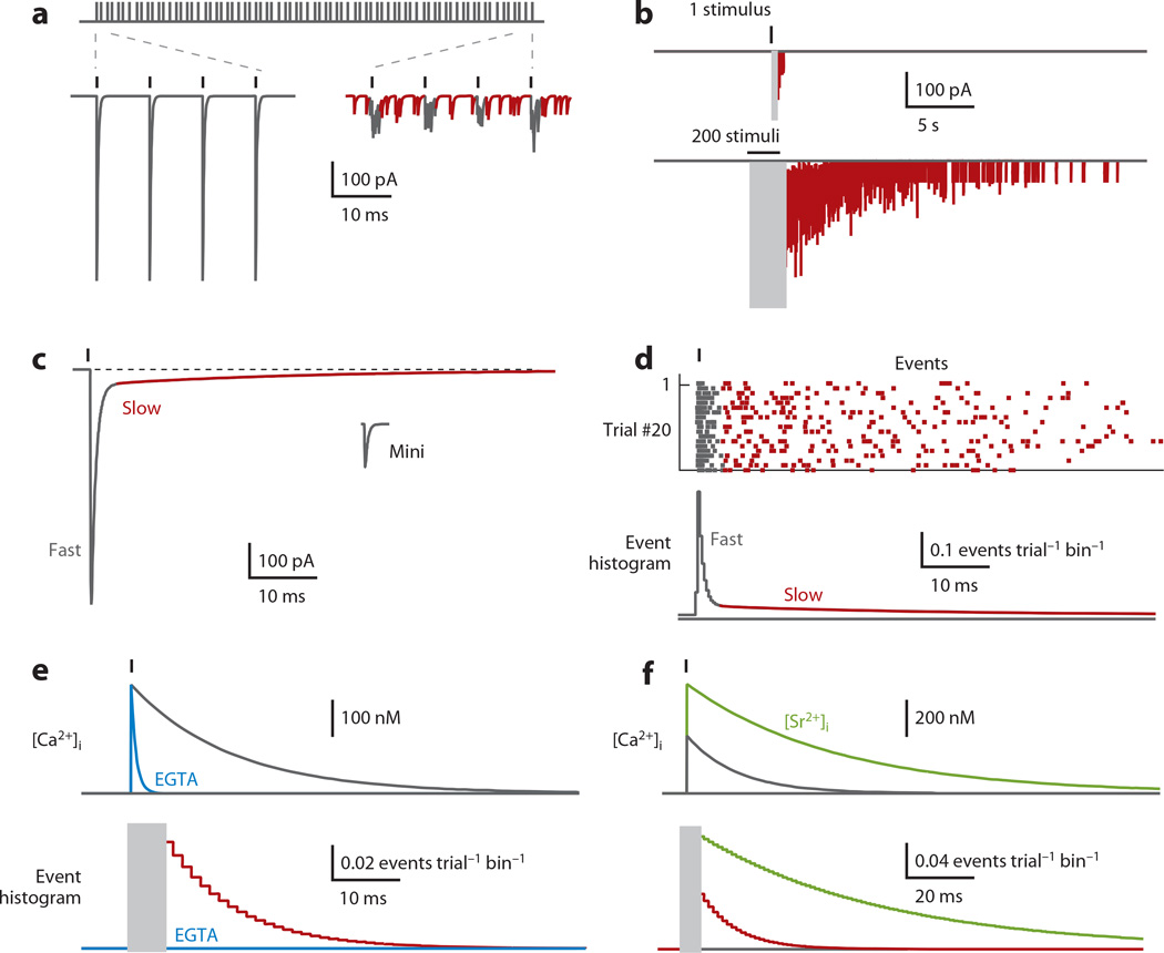
Most neuronal communication relies upon the synchronous release of neurotransmitters, which occurs through synaptic vesicle exocytosis triggered by action potential invasion of a presynaptic bouton. However, neurotransmitters are also released asynchronously with a longer, variable delay following an action potential or spontaneously in the absence of action potentials. A compelling body of research has identified roles and mechanisms for synchronous release, but asynchronous release and spontaneous release are less well understood. In this review, we analyze how the mechanisms of the three release modes overlap and what molecular pathways underlie asynchronous and spontaneous release. We conclude that the modes of release have key fusion processes in common but may differ in the source of and necessity for Ca(2+) to trigger release and in the identity of the Ca(2+) sensor for release.


Figures

Figure 1
Figure 1
Different types of synaptic transmission illustrated with simulated data. (a) Stimulation (arrowhead) evokes synchronous and asynchronous release. (b) Spontaneous neurotransmitter release is shown on a different timescale. The inset shows a miniature postsynaptic current on an expanded timescale. Abbreviations: mPSC, miniature PSC; PSC, postsynaptic current.
Figure 2
Figure 2
Precision of synchronous release relies on brief Ca2+ channel signals and on the rapid kinetics of the Ca2+ sensor. The schematic illustrates the rapid depolarization and repolarization of an action potential, and the speed of the resulting Ca2+ current (ICa) and vesicle fusion.
Figure 3
Figure 3
Schematic of mechanisms for synchronous release of neurotransmitters at central nervous synapses. The four mechanisms are (a) tethering of synaptic vesicles close to presynaptic, voltage-gated Ca2+ channels at specialized sites for release; (b) rendering vesicles release ready during priming under the control of active zone molecules Munc13 and RIM, which may involve partial assembly of SNARE complexes; (c) action potential–induced, brief opening of voltage-gated Ca2+ channels to allow for a sharp rise and decay of Ca2+ near vesicles; and (d) fast binding of Ca2+ to the synchronous Ca2+ sensor synaptotagmin 1 to trigger fusion of synaptic vesicles. The sensor also needs to have a fast off-rate for limiting high release rates to a few hundred microseconds. These steps synchronize release, and asynchronous release and spontaneous release may mechanistically differ in one of these four steps. Abbreviations: Cpx, complexin; ELKS, protein rich in the amino acids E, L, K, and S; RIM, Rab3-interacting molecule; RIM-BP, RIM-binding protein; Stx, syntaxin-1; Syb, synaptobrevin 2 (also termed VAMP2); Syt1/2/9, synaptotagmin 1, 2, or 9.
Figure 4
Figure 4
Properties of asynchronous release. Schematics and simulated data are used to illustrate properties of asynchronous release (indicated by red). (a) At some synapses, sustained high-frequency activation initially evokes synchronous release (left), but after prolonged stimulation release is desynchronized (right). (b) At many synapses, a single stimulation evokes asynchronous release lasting tens of milliseconds (upper), whereas prolonged high-frequency stimulation produces asynchronous release lasting tens of seconds (lower). Large currents corresponding to synchronous release are blanked (gray boxes). (c) In some cases, average synaptic currents are used to estimate the time course of neurotransmitter release, and a slow component is used to estimate the amplitude and duration of asynchronous release. An average miniature synaptic current, during which all the transmitter is released at the same time, is shown for comparison. (d) Ideally, asynchronous release is characterized as individual quantal events, as illustrated by an example in which 20 trials (upper) are displayed in an event histogram of detected events (lower). (e) The slow Ca2+ buffer EGTA has been used to demonstrate that asynchronous release is Ca2+ dependent. EGTA has little effect on peak Ca2+ levels (upper) or synchronous release (lower; blanked out by gray box), but it chelates Ca2+ to abolish asynchronous release (lower blue), which is present in the absence of EGTA (lower red). (f) Replacing extracellular Ca2+ with Sr2+ increases the amplitude and duration of asynchronous release (lower green). This is primarily a consequence of Sr2+ being less well buffered and extruded by presynaptic boutons, which results in larger, longer-lasting Sr2+ signals (upper green) that drive release via the machinery for synchronous release.
Figure 5
Figure 5
The Ca2+ dependence of neurotransmitter release. The dependence of release on intracellular Ca2+ was established at the calyx of Held in wild-type (WT) animals and in knockout (ko) animals in which the fast Ca2+ sensor synaptotagmin 2 (Syt2) has been eliminated. In wild-type animals, a steep power law dependence is seen for Ca2+ levels above 0.5 µM (n= 5), and for lower Ca2+ levels there is a much shallower Ca2+ dependence (n= 2). In Syt2 ko animals, release for all ranges of Ca2+ is approximated by n= 2. A dashed line shows the Ca2+ dependence as a function of release for a sensor with n= 5. These findings indicate that, at the calyx of Held, a Ca2+ sensor other than Syt2 dominates release when Ca2+ levels are less than ~0.5 µM in WT animals. Simulated data inspired by Reference 67.
Figure 6
Figure 6
Schematic of proposed points of divergence of the modes of neurotransmitter release. Several mechanisms may be involved in promoting asynchronous and/or spontaneous release as opposed to synchronous release. Top graphs depict a presynaptic nerve terminal filled with synaptic vesicles. Bottom graphs show an expanded schematic of the presynaptic plasma membrane and a synaptic vesicle to highlight potential mechanisms. (a) Asynchronous or spontaneous release may be driven by distinct sources of Ca2+; potential sources include Ca2+ currents through Ca2+-permeable TRPV1 channels or P2×2 purinergic receptors, slow or stochastic currents through CaV2 voltage-gated Ca2+ channels, bulk cytosolic or resting Ca2+, and Ca2+ from internal stores. (b) Divergent Ca2+ sensors may control properties of release. Fast synaptotagmins (Syt1, −2, and −9) trigger synchronous release. Additional Ca2+ sensors may be involved in mediating asynchronous or spontaneous release. Synaptotagmin 7 (Syt7) was originally proposed to be a potential Ca2+ sensor that may be localized on the presynaptic plasma membrane. Recent experiments delivered arguments for and against Syt7 operating as a Ca2+ sensor for asynchronous release (Table 1). Doc2 proteins are cytosolic C2-domain proteins that were recently promoted as Ca2+ sensors for spontaneous or asynchronous release, but the data are controversial (Tables 1 and 2). (c) Distinct vesicle pools may also be involved in the mechanisms of spontaneous and asynchronous release. In particular, alternative vesicular SNARE proteins (Vti1a, VAMP7, and VAMP4) have been proposed to be associated with vesicles that specifically promote asynchronous or spontaneous release (Table 2).

References

    1. Katz B, Miledi R. The measurement of synaptic delay, and the time course of acetylcholine release at the neuromuscular junction. Proc. R. Soc. Lond. B. 1965;161:483–495. - PubMed
    1. Borst JG, Sakmann B. Calcium influx and transmitter release in a fast CNS synapse. Nature. 1996;383:431–434. - PubMed
    1. Sabatini BL, Regehr WG. Timing of neurotransmission at fast synapses in the mammalian brain. Nature. 1996;384:170–172. - PubMed
    1. Llinás R, Steinberg IZ, Walton K. Presynaptic calcium currents in squid giant synapse. Biophys. J. 1981;33:289–321. - PMC - PubMed
    1. Chen C, Regehr WG. Contributions of residual calcium to fast synaptic transmission. J. Neurosci. 1999;19:6257–6266. - PMC - PubMed

Publication types

LinkOut - more resources