Review
. 2013:47:139-65.
doi: 10.1146/annurev-genet-111212-133424.

Organizing principles of mammalian nonsense-mediated mRNA decay

Affiliations
Review

Organizing principles of mammalian nonsense-mediated mRNA decay

Maximilian Wei-Lin Popp et al. Annu Rev Genet. 2013.

Abstract

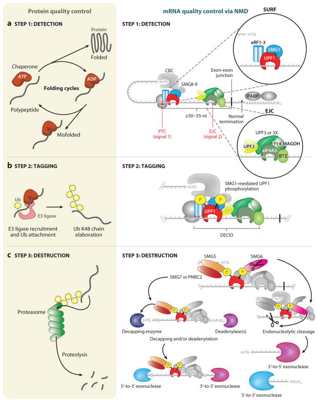
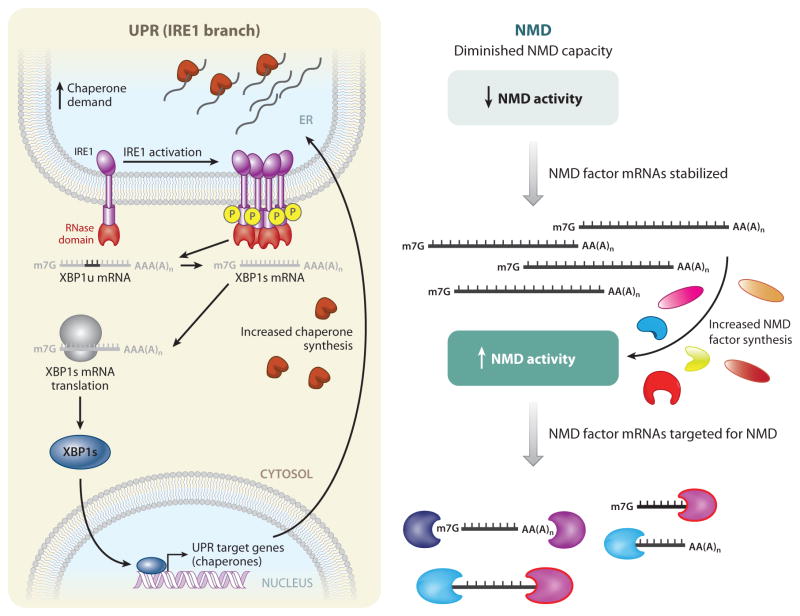
Cells use messenger RNAs (mRNAs) to ensure the accurate dissemination of genetic information encoded by DNA. Given that mRNAs largely direct the synthesis of a critical effector of cellular phenotype, i.e., proteins, tight regulation of both the quality and quantity of mRNA is a prerequisite for effective cellular homeostasis. Here, we review nonsense-mediated mRNA decay (NMD), which is the best-characterized posttranscriptional quality control mechanism that cells have evolved in their cytoplasm to ensure transcriptome fidelity. We use protein quality control as a conceptual framework to organize what is known about NMD, highlighting overarching similarities between these two polymer quality control pathways, where the protein quality control and NMD pathways intersect, and how protein quality control can suggest new avenues for research into mRNA quality control.


Figures

Figure 1
Figure 1
Similar steps govern protein quality control and nonsense-mediated mRNA decay (NMD). A general overview of protein quality control (left) and mRNA quality control, as exemplified by NMD (right), can be separated into three distinct steps: detection, tagging, and destruction. (a) Detection for protein quality control proceeds through ATP-dependent cycles of chaperone binding, release, and rebinding to hydrophobic patches in unfolded and/or partially folded proteins, providing the opportunity to fold (left). A decision is made as to whether the client protein is terminally misfolded, and, if so, this leads to tagging. The detection step for NMD relies on two signals (right). The first signal is provided by the premature termination codon (PTC), which generally defines NMD substrates, and the proteins that associate with the translation termination complex that assembles at the PTC, including the terminating ribosome and the SURF complex (inset, upper right), which consists of SMG1, UPF1, eRF1, and eRF3. The second signal often derives from an exon-junction complex (EJC), which associates with mRNAs <24 nucleotides (nts) upstream of a splicing-generated exon-exon junction whether or not they are NMD substrates. Other signals, such as unusually long 3′ UTRs, also exist. The EJC (inset, lower right) is composed of four core components (eIF4A3, Y14, MAGOH, BTZ) and associated NMD factors UPF3 or UPF3X and UPF2. If a PTC is located ≥50–55 nts upstream of an exon-exon junction, the mRNA is recognized as aberrant and tagging proceeds. (b) (Left) Tagging for misfolded proteins is governed by the ubiquitylation system. In an ATP-dependent reaction, ubiquitin (Ub) is covalently transferred from an E1 enzyme to an E2 enzyme (not shown). Ub can be transferred in covalent linkage to E3 enzymes containing a HECT (homologous to the E6-AP carboxyl terminus) domain before transfer to the substrate protein, or Ub can be directly transferred from the E2 enzyme to substrates by E3 ligase enzymes containing a RING (really interesting new gene) domain. E3 ligases can mediate ubiquitin transfer to misfolded proteins by binding to molecular chaperones. Mono-Ub is elaborated into a Ub chain with linkages at lysine (K)48. This constitutes the tag that identifies the attached protein for destruction. (Right) For tagging during NMD to occur, all or some of the SURF complex joins the EJC, possibly while the terminating ribosome is still present, forming a decay-inducing complex (DECID). This configuration activates SMG1 to phosphorylate UPF1. Phosphorylated UPF1 signals the mRNA for destruction and has the added effect of inducing translational repression of the mRNA, which is a prerequisite for destruction. (c) (Left) Destruction of proteins is the job of the proteasome, a macromolecular proteolytic machine whose components include proteins that recognize Ub-chain tags and initiate feeding of the aberrant protein into the bore of the proteasome. Aberrant proteins are thereby degraded into short peptides. (Right) NMD-dependent destruction of mRNA relies on recruitment of SMG6 and/or SMG5-SMG7 or SMG5 -PNRC2 complexes via the phosphate tags on UPF1. SMG6 possesses its own endonucleolytic cleavage activity, cutting the mRNA target 5′ to the EJC. 3′-to-5′ exonucleases degrade the 5′-cleavage product. UPF1 helicase activity disassembles the RNP components bound to the 3′-cleavage product, and this is followed by 5′-to-3′ exonucleolytic degradation. The SMG5-SMG7 or SMG5-PNRC2 adaptor complexes, recruited via SMG5 to a UPF1-localized phosphate moiety, direct exonucleolytic degradation of the mRNA. These adaptors recruit decapping enzymes and/or deadenylation enzymes, and their activities are followed by 5′-to-3′ and 3′-to-5′ exonucleolytic decay, respectively. Proteins relevant to each step are shown in color. Abbreviation: CBC, cap-binding complex.
Figure 2
Figure 2
Regulation of protein quality control and nonsense-mediated mRNA decay (NMD) provides buffering capacity. Protein quality control (left), as exemplified by the IRE1-mediated branch of the unfolded protein response (UPR), and mRNA quality control, as exemplified by NMD (right), are both governed by regulatory loops that provide buffering capacity. (Left) For IRE1-mediated UPR activation, increased demand on the protein-folding load of the endoplasmic reticulum (ER) leads to an accumulation of unfolded and/or partially folded proteins and an increased demand for more folding chaperones. IRE1, a membrane-embedded sensor of unfolded proteins, is activated either by direct binding to unfolded proteins or by titration of chaperones (which prevent IRE1 oligomerization) away from IRE1. IRE1 subsequently oligomerizes and autophosphorylates. The RNase domain in activated IRE1 (red ) then mediates an unconventional cytoplasmic splicing reaction to generate spliced XBP1 (XBP1s) mRNA from unspliced (XBP1u) mRNA. XBP1s mRNA is translated into XBP1s protein, which is a transcriptional activator that upregulates a suite of UPR target genes. Among these genes are chaperones, leading to increased chaperone synthesis so as to match the protein-folding capacity of the ER with cellular demand. (Right) Some NMD factors (UPF1, UPF2, UPF3X, SMG1, SMG5, SMG6, and SMG7) are themselves normally targets of NMD by virtue of the presence of an unusually long 3′ UTR, a uORF, or both. When genetic insults diminish NMD capacity, NMD activity is decreased, and the mRNAs encoding the NMD factors are stabilized. These stabilized mRNAs direct protein synthesis in an effort to restore normal NMD activity.

References

    1. Amrani N, Ganesan R, Kervestin S, Mangus DA, Ghosh S, Jacobson A. A faux 3′-UTR promotes aberrant termination and triggers nonsense-mediated mRNA decay. Nature. 2004;432:112–18. - PubMed
    1. Arias-Palomo E, Yamashita A, Fernandez IS, Núñez-Ramirez R, Bamba Y, et al. The nonsense-mediated mRNA decay SMG-1 kinase is regulated by large-scale conformational changes controlled by SMG-8. Genes Dev. 2011;25:153–64. - PMC - PubMed
    1. Ballinger CA, Connell P, Wu Y, Hu Z, Thompson LJ, et al. Identification of CHiP, a novel tetra-tricopeptide repeat-containing protein that interacts with heat shock proteins and negatively regulates chaperone functions. Mol Cell Biol. 1999;19:4535–45. - PMC - PubMed
    1. Ballut L, Marchadier B, Baguet A, Tomasetto C, Séraphin B, Le Hir H. The exon junction core complex is locked onto RNA by inhibition of eIF4AIII ATPase activity. Nat Struct Mol Biol. 2005;12:861–69. - PubMed
    1. Belgrader P, Cheng J, Zhou X, Stephenson LS, Maquat LE. Mammalian nonsense codons can be cis effectors of nuclear mRNA half-life. Mol Cell Biol. 1994;14:8219–28. - PMC - PubMed

Publication types

MeSH terms