

Biology and genetics of prions causing neurodegeneration
- PMID: 24274755
- PMCID: PMC4010318
- DOI: 10.1146/annurev-genet-110711-155524
Biology and genetics of prions causing neurodegeneration
Abstract
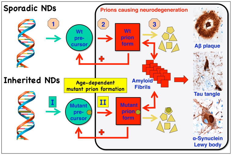
Prions are proteins that acquire alternative conformations that become self-propagating. Transformation of proteins into prions is generally accompanied by an increase in β-sheet structure and a propensity to aggregate into oligomers. Some prions are beneficial and perform cellular functions, whereas others cause neurodegeneration. In mammals, more than a dozen proteins that become prions have been identified, and a similar number has been found in fungi. In both mammals and fungi, variations in the prion conformation encipher the biological properties of distinct prion strains. Increasing evidence argues that prions cause many neurodegenerative diseases (NDs), including Alzheimer's, Parkinson's, Creutzfeldt-Jakob, and Lou Gehrig's diseases, as well as the tauopathies. The majority of NDs are sporadic, and 10% to 20% are inherited. The late onset of heritable NDs, like their sporadic counterparts, may reflect the stochastic nature of prion formation; the pathogenesis of such illnesses seems to require prion accumulation to exceed some critical threshold before neurological dysfunction manifests.
Figures
References
-
- Alper T, Haig DA, Clarke MC. The exceptionally small size of the scrapie agent. Biochem Biophys Res Commun. 1966;22:278–84. - PubMed
-
- Alzheimer A. Ueber eine eigenartige Erkrankung der Hirnrinde. Cent Nervenheilk Psychiat. 1907;30:177–79.
-
- Baker HF, Ridley RM, Duchen LW, Crow TJ, Bruton CJ. Induction of β(A4)-amyloid in primates by injection of Alzheimer’s disease brain homogenate. Mol Neurobiol. 1994;8:25–39. - PubMed
-
- Baskakov IV, Legname G, Baldwin MA, Prusiner SB, Cohen FE. Pathway complexity of prion protein assembly into amyloid. J Biol Chem. 2002;277:21140–48. - PubMed
-
- Bellinger-Kawahara CG, Kempner E, Groth DF, Gabizon R, Prusiner SB. Scrapie prion liposomes and rods exhibit target sizes of 55,000 Da. Virology. 1988;164:537–41. - PubMed
Publication types
MeSH terms
Substances
Grants and funding
LinkOut - more resources
Full Text Sources
Other Literature Sources
Medical
Molecular Biology Databases
