. 2014 Jan 10;289(2):895-908.
doi: 10.1074/jbc.M113.507913. Epub 2013 Nov 25.

A direct interaction between leucine-rich repeat kinase 2 and specific β-tubulin isoforms regulates tubulin acetylation

Affiliations

A direct interaction between leucine-rich repeat kinase 2 and specific β-tubulin isoforms regulates tubulin acetylation

Bernard M H Law et al. J Biol Chem. .

Abstract

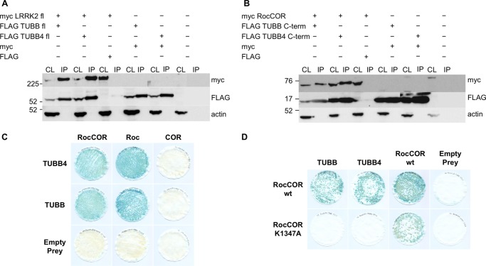
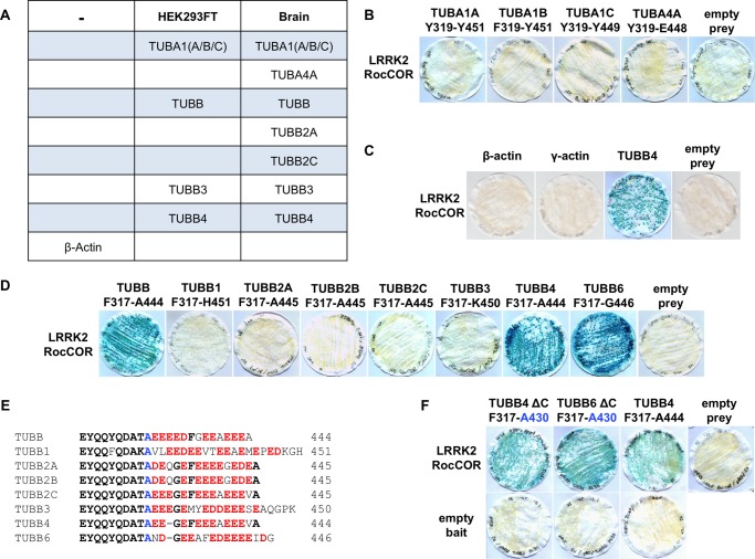
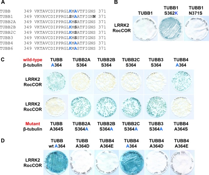
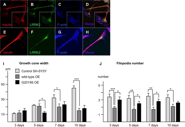
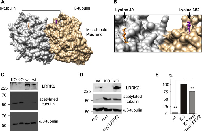
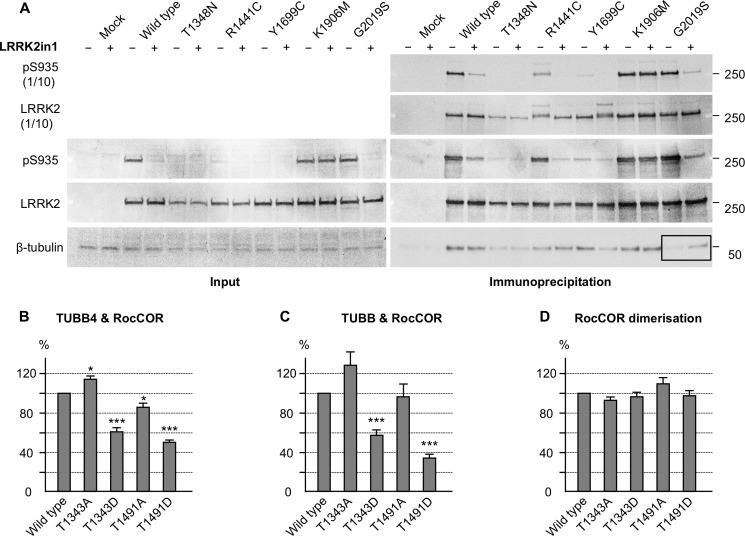
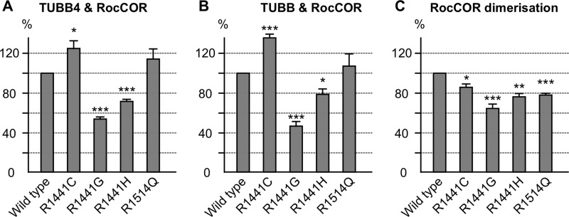
Mutations in LRRK2, encoding the multifunctional protein leucine-rich repeat kinase 2 (LRRK2), are a common cause of Parkinson disease. LRRK2 has been suggested to influence the cytoskeleton as LRRK2 mutants reduce neurite outgrowth and cause an accumulation of hyperphosphorylated Tau. This might cause alterations in the dynamic instability of microtubules suggested to contribute to the pathogenesis of Parkinson disease. Here, we describe a direct interaction between LRRK2 and β-tubulin. This interaction is conferred by the LRRK2 Roc domain and is disrupted by the familial R1441G mutation and artificial Roc domain mutations that mimic autophosphorylation. LRRK2 selectively interacts with three β-tubulin isoforms: TUBB, TUBB4, and TUBB6, one of which (TUBB4) is mutated in the movement disorder dystonia type 4 (DYT4). Binding specificity is determined by lysine 362 and alanine 364 of β-tubulin. Molecular modeling was used to map the interaction surface to the luminal face of microtubule protofibrils in close proximity to the lysine 40 acetylation site in α-tubulin. This location is predicted to be poorly accessible within mature stabilized microtubules, but exposed in dynamic microtubule populations. Consistent with this finding, endogenous LRRK2 displays a preferential localization to dynamic microtubules within growth cones, rather than adjacent axonal microtubule bundles. This interaction is functionally relevant to microtubule dynamics, as mouse embryonic fibroblasts derived from LRRK2 knock-out mice display increased microtubule acetylation. Taken together, our data shed light on the nature of the LRRK2-tubulin interaction, and indicate that alterations in microtubule stability caused by changes in LRRK2 might contribute to the pathogenesis of Parkinson disease.

Keywords: Cytoskeletal Dynamics; GTPase Mutation; Growth Cone; Lrrk2; Microtubules; Molecular Genetics; Parkinson Disease; RocCOR; Tubulin; Tubulin Acetylation.


Figures

FIGURE 1.
FIGURE 1.
The LRRK2 Roc GTPase domain binds TUBB and TUBB4. A, full-length myc-tagged LRRK2 interacts with full-length FLAG-tagged TUBB and TUBB4 in co-immunoprecipitation experiments (CL, cell lysate; IP, = anti-FLAG immunoprecipitates; transfections as indicated). B, the LRRK2 RocCOR domain and the C termini of interacting β-tubulin isoforms were sufficient for interaction. C, semiquantitative YTH experiments show interaction with TUBB and TUBB4 to require the LRRK2 Roc but not COR domain. D, semiquantitative lacZ freeze-fracture assays show that the LRRK2 guanine nucleotide-binding mutant K1347A abolishes LRRK2 interactions with TUBB and TUBB4 but not LRRK2 RocCOR dimerization.
FIGURE 2.
FIGURE 2.
The direct interaction between LRRK2 and tubulin is specific to TUBB, TUBB4, and TUBB6. A, mass spectrometry of GST-Roc immunoprecipitation from brain and HEK293 cell lysates identified α-tubulin and β-tubulin isoforms but not β-actin as LRRK2 interactors. B, YTH assays show that the LRRK2 RocCOR tandem domain does not bind directly to the α-tubulin isoforms TUBA1A, TUBA1B, TUBA1C, and TUBA4A; or C, β- or γ-actin. D, LRRK2 binds directly to the C termini of TUBB, TUBB4, and TUBB6 but not to TUBB1, TUBB2A-C, or TUBB3. E, sequence alignment of the divergent extreme C-terminal tails of all eight β-tubulins. F, deletion of these C-terminal amino acid residues does not abolish the interaction between LRRK2 and β-tubulin in YTH assays.
FIGURE 3.
FIGURE 3.
The LRRK2 β-tubulin interaction requires lysine 362 and alanine 364 of β-tubulin. A, alignment of β-tubulin isoforms reveals further sequence divergence between residues 349 and 371. Note the presence of Lys-362 and Ala-364 in isoforms found to bind LRRK2 (TUBB, TUBB4, and TUBB6). B, mutation S362K allows TUBB1 to bind LRRK2. C, mutation A364S in TUBB and TUBB4 abolishes the interaction of these proteins with LRRK2. By contrast, the reciprocal S364A mutation in TUBB2A, TUBB2B, TUBB2C, and TUBB3 allows these β-tubulins to interact with LRRK2. D, interaction of TUBB and TUBB4 with the LRRK2 RocCOR tandem domain is also abolished by phosphomimetic mutations at residue 364.
FIGURE 4.
FIGURE 4.
Lysine 362 and alanine 364 of β-tubulin change the conformation of the LRRK2 binding site. A, modeling shows the LRRK2 and taxol binding sites are in close proximity at the luminal surface of MTs. B, magnification of the area indicated in A. C-E, modeling of the structural influence of Lys-362 and Ala-364 versus Ser-362 and Ser-364 on the accessibility of the LRRK2 binding site. C, Ala-364 in TUBB and TUBB4 is unlikely to form hydrogen bonds with arginine 318 allowing for good accessibility of Lys-362 at the MT surface. D, by contrast, Ser-364 is predicted to form a hydrogen bond with arginine 318 restricting Lys-362 conformation. E, the shorter side chain of Ser-362 in comparison to Lys-362 at the MT surface is predicted to be unable to coordinate LRRK2 binding. A and B are derived from the crystal structure of bovine α/β-tubulin dimers bound to taxol; C–E are derived from the crystal structure of unbound porcine α/β-tubulin dimers.
FIGURE 5.
FIGURE 5.
Mutations to TUBB6 confirm the influence of arginine versus proline at residue 320 on interaction with LRRK2. A, the TUBB6 single mutants P320R, K362S, or A364S do not abolish the LRRK2 interaction with tubulin. Double mutants P320R/K362S or P320R/A364S in TUBB6 abolish the interaction with LRRK2. B, alignment of β-tubulin isoforms reveals sequence divergence between TUBB6 and all other β-tubulins at residue 320. C, modeling shows Arg-320 in β-tubulin in close proximity to the LRRK2 binding residue Lys-362. D, magnification of the area indicated in C. E, modeling of the structural influence of Arg-320 versus Pro-320 on the accessibility of the LRRK2 binding site.
FIGURE 6.
FIGURE 6.
LRRK2 expression levels and the G2019S mutation affect growth cone dynamics. A–H, endogenous (A–D) and stably overexpressed (E–H) LRRK2 localizes preferentially to growth cones in SH-SY5Y neurites. Note the decline in expression levels proximal of the growth cones along the neurites. Scale bar 10 μm. I, the increase in growth cone width during differentiation observed in control cells is abolished in cells over-expressing wild-type or G2019S mutant LRRK2. J, the number of filopodia per growth cone is decreased significantly in cells over-expressing wild-type LRRK2 in comparison to control cells. By contrast, the number of filopodia per growth cone is increased significantly in cells over-expressing G2019S mutant LRRK2 in comparison to wild-type over-expressing cells. *, p < 0.05; **, p < 0.01; ***, p < 0.001; n = 10.
FIGURE 7.
FIGURE 7.
LRRK2 expressions correlates inversely with tubulin acetylation. A, modeling shows the LRRK2 binding site in close proximity to the Lys-40 acetylation site in α-tubulin at the luminal surface of MTs. B, magnification of the area indicated in Fig. 6A. C, acetylation of Lys-40 in α-tubulin is massively increased in LRRK2 knock-out MEFs. D and E, the increase in acetylation in LRRK2 knock-out MEFs can be rescued by over-expression of human LRRK2. *, p < 0.05; **, p < 0.01; ***, p < 0.001; n = 3.
FIGURE 8.
FIGURE 8.
LRRK2 Roc domain autophosphorylation interferes with the LRRK2-tubulin interaction. A, Western blot showing a decrease of the LRRK2-β-tubulin interaction in co-immunoprecipitation experiments with the G2019S mutant in comparison with wild-type LRRK2 (black box). This decrease was rescued by LRRK2 kinase inhibition with LRRK2in1. B–D, quantitative YTH assays show that the introduction of phosphomimetic mutations at Roc domain autophosphorylation sites decreases the interaction with TUBB4 (B) and TUBB (C) but has no effect on RocCOR dimerization (D). Introduction of an alanine at the autophosphorylation sites has less and more diverse effects on the interactions shown. *, p < 0.05; **, p < 0.01; ***, p < 0.001; n = 5–6.
FIGURE 9.
FIGURE 9.
Familial LRRK2 mutations affect the LRRK2 β-tubulin interaction. A and B, quantitative YTH assays show that the introduction of familial LRRK2 mutations can increase (R1441C) as well as decrease (R1441G/H) the interaction with TUBB4 (A) and TUBB (B), whereas the R1514Q amino acid change, which does not segregate with PD, has no statistically significant effect. C, all amino acid changes decrease the RocCOR dimerization strength. *, p < 0.05; **, p < 0.01; ***, p < 0.001; n = 4.

References

    1. Paisán-Ruíz C., Jain S., Evans E. W., Gilks W. P., Simón J., van der Brug M., López de Munain A., Aparicio S., Gil A. M., Khan N., Johnson J., Martinez J. R., Nicholl D., Carrera I. M., Pena A. S., de Silva R., Lees A., Martí-Massó J. F., Pérez-Tur J., Wood N. W., Singleton A. B. (2004) Cloning of the gene containing mutations that cause PARK8-linked Parkinson's disease. Neuron 44, 595–600 - PubMed
    1. Zimprich A., Biskup S., Leitner P., Lichtner P., Farrer M., Lincoln S., Kachergus J., Hulihan M., Uitti R. J., Calne D. B., Stoessl A. J., Pfeiffer R. F., Patenge N., Carbajal I. C., Vieregge P., Asmus F., Müller-Myhsok B., Dickson D. W., Meitinger T., Strom T. M., Wszolek Z. K., Gasser T. (2004) Mutations in LRRK2 cause autosomal-dominant parkinsonism with pleomorphic pathology. Neuron 44, 601–607 - PubMed
    1. Ross O. A., Toft M., Whittle A. J., Johnson J. L., Papapetropoulos S., Mash D. C., Litvan I., Gordon M. F., Wszolek Z. K., Farrer M. J., Dickson D. W. (2006) Lrrk2 and Lewy body disease. Ann. Neurol. 59, 388–393 - PubMed
    1. Kumari U., Tan E. K. (2009) LRRK2 in Parkinson's disease. Genetic and clinical studies from patients. FEBS J. 276, 6455–6463 - PubMed
    1. Berwick D. C., Harvey K. (2011) LRRK2 signaling pathways. The key to unlocking neurodegeneration? Trends Cell Biol. 21, 257–265 - PubMed

Publication types

MeSH terms