

Diagnostic value of knee arthrometry in the prediction of anterior cruciate ligament strain during landing
- PMID: 24275863
- PMCID: PMC3928813
- DOI: 10.1177/0363546513509961
Diagnostic value of knee arthrometry in the prediction of anterior cruciate ligament strain during landing
Abstract
Background: Previous studies have indicated that higher knee joint laxity may be indicative of an increased risk of anterior cruciate ligament (ACL) injuries. Despite the frequent clinical use of knee arthrometry in the evaluation of knee laxity, little data exist to correlate instrumented laxity measures and ACL strain during dynamic high-risk activities. Purpose/
Hypotheses: The purpose of this study was to evaluate the relationships between ACL strain and anterior knee laxity measurements using arthrometry during both a drawer test and simulated bipedal landing (as an identified high-risk injurious task). We hypothesized that a high correlation exists between dynamic ACL strain and passive arthrometry displacement. The secondary hypothesis was that anterior knee laxity quantified by knee arthrometry is a valid predictor of injury risk such that specimens with greater anterior knee laxity would demonstrate increased levels of peak ACL strain during landing.
Study design: Controlled laboratory study.
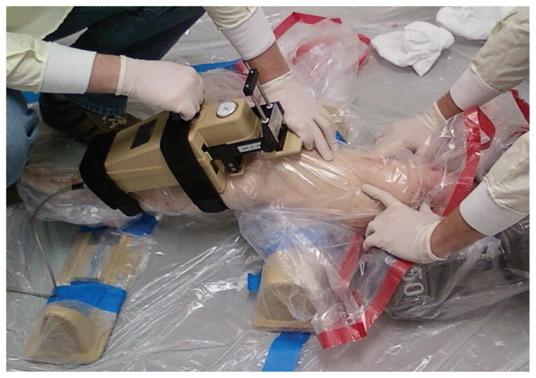
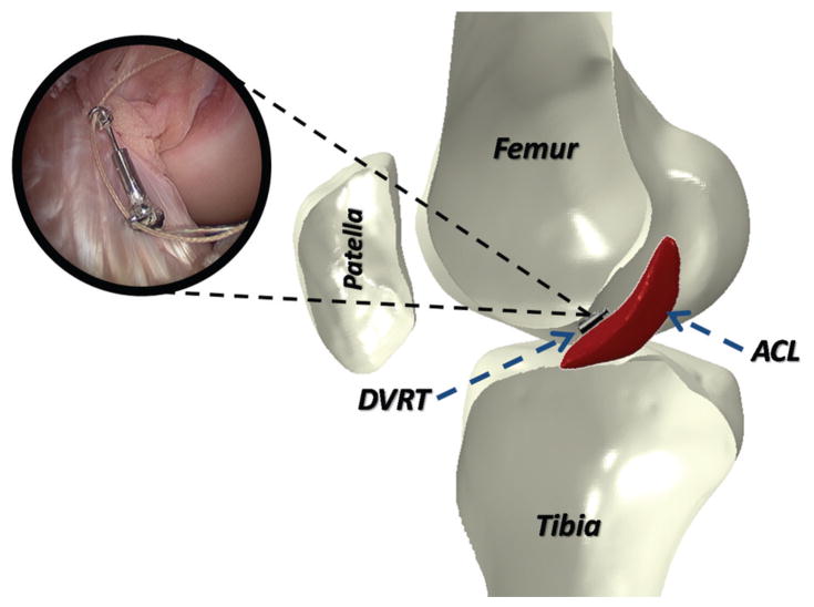
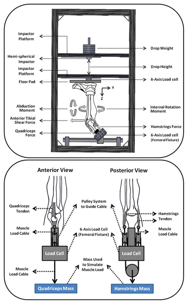
Methods: Twenty cadaveric lower limbs (mean age, 46 ± 6 years; 10 female and 10 male) were tested using a CompuKT knee arthrometer to measure knee joint laxity. Each specimen was tested under 4 continuous cycles of anterior-posterior shear force (±134 N) applied to the tibial tubercle. To quantify ACL strain, a differential variable reluctance transducer (DVRT) was arthroscopically placed on the ACL (anteromedial bundle), and specimens were retested. Subsequently, bipedal landing from 30 cm was simulated in a subset of 14 specimens (mean age, 45 ± 6 years; 6 female and 8 male) using a novel custom-designed drop stand. Changes in joint laxity and ACL strain under applied anterior shear force were statistically analyzed using paired sample t tests and analysis of variance. Multiple linear regression analyses were conducted to determine the relationship between anterior shear force, anterior tibial translation, and ACL strain.
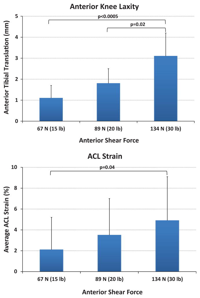
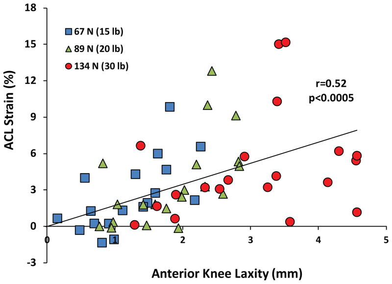
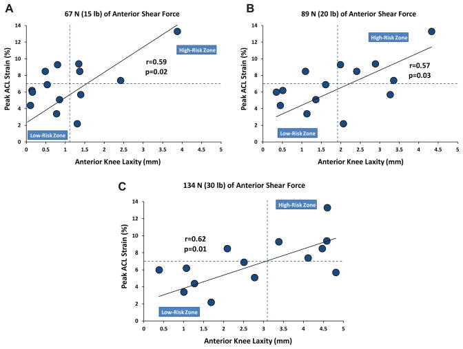
Results: During simulated drawer tests, 134 N of applied anterior shear load produced a mean peak anterior tibial translation of 3.1 ± 1.1 mm and a mean peak ACL strain of 4.9% ± 4.3%. Anterior shear load was a significant determinant of anterior tibial translation (P < .0005) and peak ACL strain (P = .04). A significant correlation (r = 0.52, P < .0005) was observed between anterior tibial translation and ACL strain. Cadaveric simulations of landing produced a mean axial impact load of 4070 ± 732 N. Simulated landing significantly increased the mean peak anterior tibial translation to 10.4 ± 3.5 mm and the mean peak ACL strain to 6.8% ± 2.8% (P < .0005) compared with the prelanding condition. Significant correlations were observed between peak ACL strain during simulated landing and anterior tibial translation quantified by knee arthrometry.
Conclusion: Our first hypothesis is supported by a significant correlation between arthrometry displacement collected during laxity tests and concurrent ACL strain calculated from DVRT measurements. Experimental findings also support our second hypothesis that instrumented measures of anterior knee laxity predict peak ACL strain during landing, while specimens with greater knee laxity demonstrated higher levels of peak ACL strain during landing.
Clinical relevance: The current findings highlight the importance of instrumented anterior knee laxity assessments as a potential indicator of the risk of ACL injuries in addition to its clinical utility in the evaluation of ACL integrity.
Keywords: ACL; arthrometry; injury; knee; laxity.
Figures
References
-
- Arms S, Boyle J, Johnson R, Pope M. Strain measurement in the medial collateral ligament of the human knee: an autopsy study. J Biomech. 1983;16(7):491–496. - PubMed
-
- Bach JM, Hull ML. Strain inhomogeneity in the anterior cruciate ligament under application of external and muscular loads. J Biomech Eng. 1998;120(4):497–503. - PubMed
-
- Boden BP, Dean GS, Feagin JA, Garrett WE. Mechanisms of anterior cruciate ligament injury. Orthopedics. 2000;23(6):573–578. - PubMed
-
- Butler DL, Noyes FR, Grood ES. Ligamentous restraints to anterior-posterior drawer in the human knee: a biomechanical study. J Bone Joint Surg Am. 1980;62(2):259–270. - PubMed
-
- Daniel DM, Malcom LL, Losse G, Stone ML, Sachs R, Burks R. Instrumented measurement of anterior laxity of the knee. J Bone Joint Surg Am. 1985;67(5):720–726. - PubMed
MeSH terms
Grants and funding
LinkOut - more resources
Full Text Sources
Other Literature Sources
