. 2013 Dec 16;210(13):2813-21.
doi: 10.1084/jem.20131244. Epub 2013 Nov 25.

Broadly neutralizing antibodies that inhibit HIV-1 cell to cell transmission

Affiliations

Broadly neutralizing antibodies that inhibit HIV-1 cell to cell transmission

Marine Malbec et al. J Exp Med. .

Abstract

The neutralizing activity of anti-HIV-1 antibodies is typically measured in assays where cell-free virions enter reporter cell lines. However, HIV-1 cell to cell transmission is a major mechanism of viral spread, and the effect of the recently described broadly neutralizing antibodies (bNAbs) on this mode of transmission remains unknown. Here we identify a subset of bNAbs that inhibit both cell-free and cell-mediated infection in primary CD4(+) lymphocytes. These antibodies target either the CD4-binding site (NIH45-46 and 3BNC60) or the glycan/V3 loop (10-1074 and PGT121) on HIV-1 gp120 and act at low concentrations by inhibiting multiple steps of viral cell to cell transmission. These antibodies accumulate at virological synapses and impair the clustering and fusion of infected and target cells and the transfer of viral material to uninfected T cells. In addition, they block viral cell to cell transmission to plasmacytoid DCs and thereby interfere with type-I IFN production. Thus, only a subset of bNAbs can efficiently prevent HIV-1 cell to cell transmission, and this property should be considered an important characteristic defining antibody potency for therapeutic or prophylactic antiviral strategies.


Figures

Figure 1.
Figure 1.
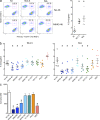
An assay for analyzing inhibition of HIV-1 cell to cell transmission by bNAbs. (a) bNAbs NIH45-46 and 8ANC195 (15 µg/ml) were incubated for 1 h with primary CD4+ T cells infected with HIV-1 (NL4.3 or NLAD8 strains). FarRed-loaded autologous target CD4+ T cells were added, and Gag+ target cells were measured 48 h later by flow cytometry. One representative experiment (out of four) is shown. The reverse transcription inhibitor NVP was used as a positive control. NI, noninfected cells. (b) Cell-associated inhibition assays were performed as in panel a. bNAbs were used at the indicated doses, and the percentage of inhibition of infection was calculated. For cell-free infections, viruses (NL4.3 or NLAD8 strains) were incubated with the indicated bNAbs for 1 h and then added to HeLa-derived P4C5 target cells, which carry an HIV-1 LTR–β-gal reported cassette. After 36 h, infection was quantified by measuring β-gal activity. Data are mean ± SD from four independent experiments. Lines represent fitted results.
Figure 2.
Figure 2.
Effect of bNAbs on formation of conjugates of infected and target cells. (a) CFSE-stained MOLT cells chronically infected with HIV-1 NL4.3 or BaL strains were preincubated for 1 h with 15 µg/ml NIH45-46 bNAb and then co-cultured with FarRed-stained primary CD4+ T cells. After 2 h, conjugates of donors and targets (CFSE+FarRed+) were quantified by flow cytometry. (left) Flow plots from one representative experiment are shown. (right) Frequency of conjugates with noninfected (NI) and NL4.3- or BaL-infected MOLT cells in the absence of bNAb. Each dot represents an experiment with primary target T cells from independent donors. The bars represent SD. *, P < 0.05. (b) Conjugate formation in the presence of the indicated bNAbs (15 µg/ml) as determined in panel a. Data are shown from six independent experiments. The percentage of cell-forming conjugates is shown. *, P < 0.05 (Wilcoxon matched pairs test). (c) HeLa cells stably expressing HIV-1 Env (NL4.3) and Tat were preincubated for 1 h with the indicated bNAbs (15 µg/ml) before overnight co-culture with HeLa P4C5 cells, which carry an HIV-1 LTR–β-gal reporter cassette. Upon syncytia formation, Tat will transactivate the HIV-1 LTR. Levels of syncytia were quantified by measuring β-gal activity. Data represent mean ± SD of triplicate samples from six independent experiments. *, P < 0.05.
Figure 3.
Figure 3.
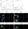
Effect of bNAbs on HIV-1 capture by target cells. (a) Primary HIV-1–infected CD4+ T cells (NL4.3 or NLAD8 strains) were preincubated for 1 h with the indicated bNAbs (15 µg/ml) before co-culture with autologous target cells stained with FarRed. After 4 h, the fraction of target cells having captured viral material (Gag+) was measured by flow cytometry. Data are mean ± SD of six independent experiments. *, P < 0.05. (b) FarRed+ HIV-1–infected CD4+ T cells or Jurkat cells were preincubated for 1 h with the indicated bNAbs before a 1.5-h co-culture with target cells stained with CFSE. Cells were stained with anti-Gag mAbs and with a secondary anti–human antibody (yellow) to visualize the bNAb. Examples of virological synapses in primary CD4+ T cells (top) and in Jurkat cells (bottom) are shown. D, donor cell; T, target cell. (c) The co-cultures of primary CD4+ T cells described in b were analyzed for target cells that were not or no longer in contact with donor cells. Images are representative of three independent experiments. Bars, 5 µm.
Figure 4.
Figure 4.
Effect of bNAbs on HIV-1 sensing by hematopoietic cells. (a) HIV-1–infected MT4C5 lymphoblastoid T cells were incubated for 1 h with the indicated bNAbs (15 µg/ml) and used as donors. Target cells were PBMCs, and levels of type-I IFN released in supernatants were measured 18 h later. NI, co-culture with noninfected cells. Data are mean ± SD of at least three independent experiments. *, P < 0.05. (b and c) HIV-infected MT4C5 cells were incubated with the indicated bNAbs for 1 h before mixing with Gen2.2 cells. After 48 h, the levels of cells productively infected (Gag+; b) or expressing the IFN-stimulated protein MxA (c) were measured by flow cytometry. Data are mean ± SD of at least three independent experiments. *, P < 0.05.

References

    1. Abela I.A., Berlinger L., Schanz M., Reynell L., Günthard H.F., Rusert P., Trkola A. 2012. Cell-cell transmission enables HIV-1 to evade inhibition by potent CD4bs directed antibodies. PLoS Pathog. 8:e1002634 10.1371/journal.ppat.1002634 - DOI - PMC - PubMed
    1. Balazs A.B., Chen J., Hong C.M., Rao D.S., Yang L., Baltimore D. 2012. Antibody-based protection against HIV infection by vectored immunoprophylaxis. Nature. 481:81–84 10.1038/nature10660 - DOI - PMC - PubMed
    1. Bonsignori M., Montefiori D.C., Wu X., Chen X., Hwang K.K., Tsao C.Y., Kozink D.M., Parks R.J., Tomaras G.D., Crump J.A., et al. 2012. Two distinct broadly neutralizing antibody specificities of different clonal lineages in a single HIV-1-infected donor: implications for vaccine design. J. Virol. 86:4688–4692 10.1128/JVI.07163-11 - DOI - PMC - PubMed
    1. Burton D.R., Ahmed R., Barouch D.H., Butera S.T., Crotty S., Godzik A., Kaufmann D.E., McElrath M.J., Nussenzweig M.C., Pulendran B., et al. 2012. A blueprint for HIV vaccine discovery. Cell Host Microbe. 12:396–407 10.1016/j.chom.2012.09.008 - DOI - PMC - PubMed
    1. Chen P., Hübner W., Spinelli M.A., Chen B.K. 2007. Predominant mode of human immunodeficiency virus transfer between T cells is mediated by sustained Env-dependent neutralization-resistant virological synapses. J. Virol. 81:12582–12595 10.1128/JVI.00381-07 - DOI - PMC - PubMed

Publication types