

Increased number of Langerhans cells in the epidermis of diabetic foot ulcers correlates with healing outcome
- PMID: 24277309
- PMCID: PMC4349345
- DOI: 10.1007/s12026-013-8474-z
Increased number of Langerhans cells in the epidermis of diabetic foot ulcers correlates with healing outcome
Abstract
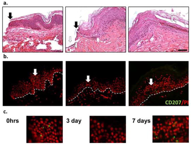
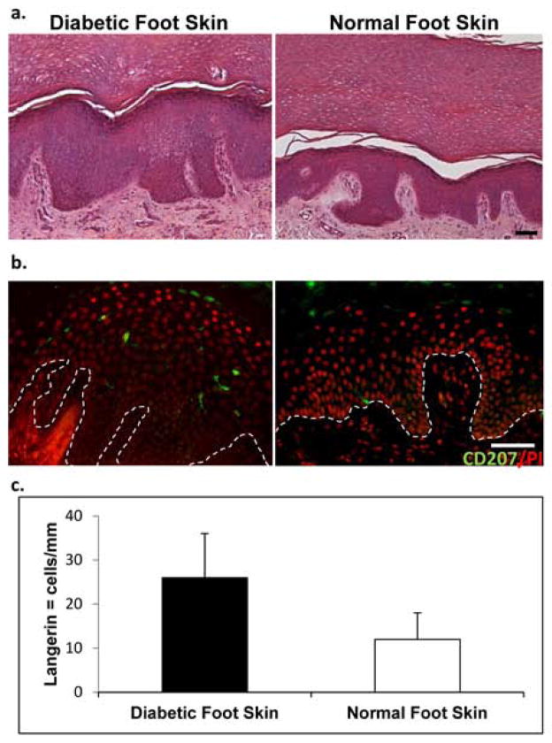
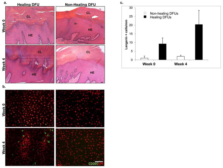
Langerhans cells (LCs) are a specialized subset of epidermal dendritic cells. They represent one of the first cells of immunologic barrier and play an important role during the inflammatory phase of acute wound healing. Despite considerable progress in our understanding of the immunopathology of diabetes mellitus and its associated comorbidities such as diabetic foot ulcers (DFUs), considerable gaps in our knowledge exist. In this study, we utilized the human ex vivo wound model and confirmed the increased epidermal LCs at wound edges during early phases of wound healing. Next, we aimed to determine differences in quantity of LCs between normal human and diabetic foot skin and to learn if the presence of LCs correlates with the healing outcome in DFUs. We utilized immunofluorescence to detect CD207+ LCs in specimens from normal and diabetic foot skin and DFU wound edges. Specimens from DFUs were collected at the initial visit and 4 weeks later at the time when the healing outcome was determined. DFUs that decreased in size by >50 % were considered to be healing, while DFUs with a size reduction of <50 % were considered non-healing. Quantitative assessment of LCs showed a higher number of LCs in healing when compared to non-healing DFU's. Our findings provide evidence that LCs are present in higher number in diabetic feet than normal foot skin. Healing DFUs show a higher number of LCs compared to non-healing DFUs. These findings indicate that the epidermal immune barrier plays an important role in the DFU healing outcome and may offer new therapeutic avenues targeting LC in non-healing DFUs.
Figures
References
-
- Hoeffel G, Wang Y, Greter M, See P, Teo P, Malleret B, et al. Adult Langerhans cells derive predominantly from embryonic fetal liver monocytes with a minor contribution of yolk sac-derived macrophages. The Journal of experimental medicine. 2012;209(6):1167–81. doi: 10.1084/jem.20120340. - DOI - PMC - PubMed
-
- Valladeau J, Ravel O, Dezutter-Dambuyant C, Moore K, Kleijmeer M, Liu Y, et al. Langerin, a novel C-type lectin specific to Langerhans cells, is an endocytic receptor that induces the formation of Birbeck granules. Immunity. 2000;12(1):71–81. - PubMed
Publication types
MeSH terms
Grants and funding
LinkOut - more resources
Full Text Sources
Other Literature Sources
Medical
