. 2014 Jan 1;192(1):523-32.
doi: 10.4049/jimmunol.1301342. Epub 2013 Nov 25.

Cancer-initiating cells from colorectal cancer patients escape from T cell-mediated immunosurveillance in vitro through membrane-bound IL-4

Affiliations

Cancer-initiating cells from colorectal cancer patients escape from T cell-mediated immunosurveillance in vitro through membrane-bound IL-4

Andrea Volonté et al. J Immunol. .

Abstract

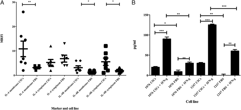
Cancer-initiating cells (CICs) that are responsible for tumor initiation, propagation, and resistance to standard therapies have been isolated from human solid tumors, including colorectal cancer (CRC). The aim of this study was to obtain an immunological profile of CRC-derived CICs and to identify CIC-associated target molecules for T cell immunotherapy. We have isolated cells with CIC properties along with their putative non-CIC autologous counterparts from human primary CRC tissues. These CICs have been shown to display "tumor-initiating/stemness" properties, including the expression of CIC-associated markers (e.g., CD44, CD24, ALDH-1, EpCAM, Lgr5), multipotency, and tumorigenicity following injection in immunodeficient mice. The immune profile of these cells was assessed by phenotype analysis and by in vitro stimulation of PBMCs with CICs as a source of Ags. CICs, compared with non-CIC counterparts, showed weak immunogenicity. This feature correlated with the expression of high levels of immunomodulatory molecules, such as IL-4, and with CIC-mediated inhibitory activity for anti-tumor T cell responses. CIC-associated IL-4 was found to be responsible for this negative function, which requires cell-to-cell contact with T lymphocytes and which is impaired by blocking IL-4 signaling. In addition, the CRC-associated Ag COA-1 was found to be expressed by CICs and to represent, in an autologous setting, a target molecule for anti-tumor T cells. Our study provides relevant information that may contribute to designing new immunotherapy protocols to target CICs in CRC patients.


Conflict of interest statement

Disclosures

The authors have no financial conflicts of interest.

Figures

FIGURE 1.
FIGURE 1.
Characterization of CIC properties in CRC sphere-forming cell cultures. The tumorigenic properties of CICs compared with autologous FBS tumor cells (A, B) were evaluated by the s.c. injection of 1 × 10⁵, 1 × 10⁴, 1 × 10³, 1 × 10², and 10 cells into immunodeficient NOD/SCID CB17 mice (see Materials and Methods). Data are indicated as the means ± SD of tumor volumes deriving from three independent experiments. Three mice per treatment were used for each experiment. *p < 0.05, **p < 0.01 (99% t test). (C) Either CICs or FBS tumor cells (1 × 10⁵ cells) were mixed at a 1:1 ratio in Matrigel-containing medium (DMEM with 1% penicillin/streptomycin; 100 μl of final volume) and transplanted into immunodeficient NOD/SCID CB17 mice. Tumor formation was monitored weekly, and when tumor sizes reached 1.5 cm3 the mice were sacrificed and tumor tissues were removed and mechanically processed to isolate single cells. These cells, derived from either CICs or FBS xenograft tumors, were cultured in vitro and subsequently transplanted into secondary mice and tumor formation was monitored weekly as well. (D) The table indicates the number of mice who developed tumors following the injection of either CICs or FBS tumor cells (patient #1076); the results are from the use of three mice for each condition for three experiments (no. of mice per condition = 9).
FIGURE 2.
FIGURE 2.
Expression or secretion of IL-4 and IL-4R by CICs and FBS tumor cell lines isolated from CRC patients. (A) IL-4 and IL-4R expression on CICs and FBS tumor cells [#1076, 1247, 111011, 14583, 1, 2, 3, 1039, 20299 and 1869 (A)] was evaluated by membrane or intracellular IF. For the mAbs used, see Materials and Methods. Data are represented as MRFI; significant values are MRFI ≥ 2. The results are the mean of three independent experiments (SD < 5). (B) Evaluation of soluble IL-4 in cell culture supernatants of CRC CICs or FBS tumor cells pretreated or not with IFN-γ (1000 IU/ml for 48 h) was performed by SearchLight Array Technology (see Materials and Methods). The data are expressed as picograms per milliliter. Results represent averages of duplicates with SD ≤ 10%. *p < 0.5, **p < 0.05, ***p < 0.001.
FIGURE 3.
FIGURE 3.
Restoration of T cell proliferation in vitro by blocking CIC-associated IL-4. PBMCs (1 × 10⁷) from CRC patients (patients #1076, 1247, and 14583) or healthy donors (for patients #1, 2, and 3) were stained with 1 μM CSFE and stimulated with PHA/Con A in the presence or not of 3-d cultured CICs or FBS tumor cell lines. The coculture of tumor cells and autologous or allogeneic PBMCs was performed in the presence or not of anti–IL-4 mAb, anti–IL-4R mAb, or both the mAbs. Then, cells were harvested and the CFSE profile was assessed by flow cytometry. Data are referred to CD3+ gated cells. Data represent the proliferation index that is the total number of divisions divided by the number of cells that went into division. Data represent the mean of the proliferation index for each tumor cell line from three independent experiments. **p < 0.05, ***p < 0.001.
FIGURE 4.
FIGURE 4.
Specific tumor reactivity of T cells from MLTCs of CRC patients. PBMCs isolated from CRC patients 1076, 1247, and 14583 were stimulated in vitro in the presence or not of the neutralizing IL-4 mAb with autologous irradiated CICs (AC) or, when available, FBS tumor cells (A, C). Following 3 wk of culture, the reactivity against autologous tumor cell lines was assessed by IFN-γ release (ELISPOT assay). Data are expressed as no. of spots per 5 × 10⁴ cells and are subtracted from the background of IFN-γ release of T cells alone. (D) The reactivity of CIC- or FBS-stimulated MLTCs against allogeneic HLA-matched EBV-B cells (1869) pulsed with the HLA-A24 epitope (SLQDLSELV, Ref. 16) or T2 cells loaded with the HLA-A2–restricted epitope (FMTRKLWDL, Ref. 16) from COA-1. The positive controls of T cell stimulation are PHA/Con A. Data are expressed as no. of spots per 5 × 10⁴ cells and subtracted from the background of IFN-γ release by T cells alone or stimulated with allogeneic target cells. Results represent averages of triplicates with SD ≤ 10%; statistical analysis of differences between means of IFN-γ released by T cells was performed by two-tailed t test. Each experiment has been repeated three times, showing consistency of the results.

References

    1. Clevers H 2011. The cancer stem cell: premises, promises and challenges. Nat. Med. 17: 313–319. - PubMed
    1. Ricci-Vitiani L, Fabrizi E, Palio E, and De Maria R. 2009. Colon cancer stem cells. J. Mol. Med. 87: 1097–1104. - PubMed
    1. Wilson BJ, Schatton T, Frank MH, and Frank NY. 2011. Colorectal cancer stem cells: biology and therapeutic implications. Curr Colorectal Cancer Rep 7: 128–135. - PMC - PubMed
    1. Bao S, Wu Q, McLendon RE, Hao Y, Shi Q, Hjelmeland AB, Dewhirst MW, Bigner DD, and Rich JN. 2006. Glioma stem cells promote radioresistance by preferential activation of the DNA damage response. Nature 444: 756–760. - PubMed
    1. Eramo A, Ricci-Vitiani L, Zeuner A, Pallini R, Lotti F, Sette G, Pilozzi E, Larocca LM, Peschle C, and De Maria R. 2006. Chemotherapy resistance of glioblastoma stem cells. Cell Death Differ. 13: 1238–1241. - PubMed

Publication types

MeSH terms

Substances