. 2013 Dec 10;110(50):20284-9.
doi: 10.1073/pnas.1315710110. Epub 2013 Nov 25.

Self-organization of axial polarity, inside-out layer pattern, and species-specific progenitor dynamics in human ES cell-derived neocortex

Affiliations

Self-organization of axial polarity, inside-out layer pattern, and species-specific progenitor dynamics in human ES cell-derived neocortex

Taisuke Kadoshima et al. Proc Natl Acad Sci U S A. .

Erratum in

  • Proc Natl Acad Sci U S A. 2014 May 20;111(20):7498

Abstract

Here, using further optimized 3D culture that allows highly selective induction and long-term growth of human ES cell (hESC)-derived cortical neuroepithelium, we demonstrate unique aspects of self-organization in human neocorticogenesis. Self-organized cortical tissue spontaneously forms a polarity along the dorsocaudal-ventrorostral axis and undergoes region-specific rolling morphogenesis that generates a semispherical structure. The neuroepithelium self-forms a multilayered structure including three neuronal zones (subplate, cortical plate, and Cajal-Retzius cell zones) and three progenitor zones (ventricular, subventricular, and intermediate zones) in the same apical-basal order as seen in the human fetal cortex in the early second trimester. In the cortical plate, late-born neurons tend to localize more basally to early-born neurons, consistent with the inside-out pattern seen in vivo. Furthermore, the outer subventricular zone contains basal progenitors that share characteristics with outer radial glia abundantly found in the human, but not mouse, fetal brain. Thus, human neocorticogenesis involves intrinsic programs that enable the emergence of complex neocortical features.

Keywords: corticogenesis; stratification.


Conflict of interest statement

The authors declare no conflict of interest.

Figures

Fig. 1.
Fig. 1.
Axial polarity in cortical NE self-organizes from hESCs. (A) hESC aggregates containing cortical NE visualized with foxg1::Venus on day 26. (B) Representative FACS analysis for foxg1::Venus+ populations. Gray, control (day 1 culture); green, day 34 culture under the new conditions. (C–J) Immunostaining of semispherical cortical structures self-formed from foxg1::venus hESCs. VZ, ventricular zone; CP, cortical plate. (K–N) Self-formation of axial polarity seen in hESC-derived cortical NE. Cortical hem-like tissues (Otx2+; M) were located in the flanking region of cortical NE on the side strong for the dorsocaudal markers Coup-TF1 (K) and Lhx2. A higher level of pErk signals (bracket) was observed on the side opposite to Coup-TF1 expression (N). Gradient and polarity of expression are indicated by triangles. Arrowhead, VZ (note that the gradients of marker expression are seen in the VZ). (O and P) Fgf8 treatment suppressed CoupTF1 and expanded the expression of the ventrorostral marker Sp8. (Scale bars, 1 mm in A; 200 μm in C–P.) Nuclear counter staining (blue), DAPI.
Fig. 2.
Fig. 2.
Asymmetric rounding morphogenesis in self-organized cortical NE. (A–I) Asymmetric progression of rounding morphogenesis of hESC-derived cortical NE. Arrows, boundary of a cortical NE domain in A and rolling epithelium in B–D. Red arrowheads, rolling epithelium in E. Red arrows, rounding movements of the NE (F–I). (J–L) Effect of the ROCK inhibitor Y-27632 on the rolling of cortical NE. (L) Attenuation of rolling morphogenesis with ROCK inhibitor. ***P < 0.001 in contingency table analysis (2 × 2) with Fisher’s exact test. Treatment group, n = 187 NE domains; control group, n = 130. (M and N) The rolling shape was preferentially observed on the side with strong expression of Otx2 and Coup-TF1 (dorsal and caudal markers). (O–Q) Adjacent formation of NE structures of cortex (Pax6+) and LGE (Gsh2+; with GAD65+ GABAergic neurons underneath) on day 35. The cortical side contacting the LGE domain was opposite to the side with strong rolling (arrow). (R) Interkinetic nuclear migration in the hESC-derived cortical NE on day 24 (two-photon imaging). Visualized with partial mixing of pax6::venus reporter hESCs with nonlabeled hESCs. Two daughter cells with both apical and basal processes were generated from an apically dividing progenitor (red dots). (Scale bars, 200 μm in A; 100 μm in B–H and J–N; 200 μm in O–Q.) Nuclear counter staining (blue), DAPI.
Fig. 3.
Fig. 3.
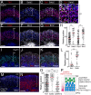
Self-formation of multiple zones in hESC-derived cortical NE. (A) Sections of day 70 hESC-derived cortical NE. Clear separation of VZ (Pax6+), SVZ, intermediate zone, and CP (Ctip2+) was seen even at this low-magnification view. (B–H) Immunostaining of day 70 cortical NE with zone-specific markers. (I) Total thickness of cortical NE and thickness of ventricular and cortical plate zones on days 70 and 91. **P < 0.01; ***P < 0.001, Student t tests between day 70 and day 91 NE samples (n = 6, each). (J–O) Immunostaining of day 91 cortical NE with zone-specific markers. (P) Schematic of the laminar structure seen in long-term culture of hESC-derived cortical NE. (Scale bars, 400 μm in A; 50 μm in B–H″; 100 μm in J–O.) Bars in graph, SEM. Nuclear counter staining (blue), DAPI.
Fig. 4.
Fig. 4.
Basally biased localization of Satb2+ and Brn2+ cortical neurons in CP. (A–H) Cortical neurons positive for Satb2 and Brn2 (superficial-layer markers) were preferentially localized to the basal (superficial) portion of the hESC-derived CP in day 91 culture. Most of the basally located Satb2+ cells were negative for the deep-layer marker Tbr1. (H) Distribution of marker-positive neurons within the CP. For relative positions, the apical and basal boundaries of the CP were defined as 0 and 100, respectively. ***P < 0.001. Mann-Whitney test. Red line, median. Counted neurons: Tbr1+ (n = 105), Satb2+ (n = 58), Ctip2+ (n = 87), and Brn2+ (n = 86). (I–L) Double-pulse labeling study using EdU (day 50; red; n = 36) and BrdU (day 70; white; n = 53). Analyzed by immunostaining on day 91. Statistical analysis was done as in H. (M–O) The mature cortical neuron marker CaMKII was preferentially expressed in Tbr1+ neurons located in the deep portion of the CP on day 112. The cortical NE was cultured on a Transwell filter during days 78–112 to support robust survival of mature neurons. (O) Plotting was done as in H. ***P < 0.001. Kruskal-Wallis test with a post hoc multiple comparison test. Numbers of neurons counted: Tbr1+ (n = 293), Satb2+ (n = 177), and CaMKII+ (n = 132). (P) Schematic of neuronal distributions within the CP of hESC-derived cortical NE on days 91 and 112. (Scale bars, 100 μm in A–C, E–G, and I–K; 50 μm in D; 200 μm in M and N.) Nuclear counter staining (blue), DAPI.
Fig. 5.
Fig. 5.
Appearance of oRG-like progenitors. (A–F) Percentages of apical progenitors with vertical (cleavage angle at 60–90°) and nonvertical (030° and 30–60°) cleavages (A and B) in the VZ of day 70 (C) and day 91 (D–F) hESC-derived cortical NE. p-Vimentin, M-phase marker. Arrowhead, pericentrin. Cells analyzed: n = 42 (day 70) and n = 33 (day 91). (G–I) Basal progenitors (Pax6+, Sox2+) and intermediate progenitors (Tbr2+) in the SVZ of day 91 culture. (H) Percentages of Sox2+/Tbr2 and Sox2/Tbr2+ progenitors within all progenitors (Sox2+ and/or Tbr2+) in the CP. The percentage of Sox2+/Tbr2 progenitors increased from day 70 to day 91, whereas Sox2/Tbr2+ progenitors decreased in proportion. ***P < 0.001, Student t tests between day 70 and day 91 samples. Non-VZ progenitors from four cortical NE domains from each day were counted. (I) On day 91, Sox2+/Tbr2 progenitors tended to localize farther from the ventricular surface than Sox2/Tbr2+ progenitors (Right). ***P < 0.001, Mann-Whitney test. Red line, median. (J–M) Pax6+ p-Vimentin+ progenitors had a long basal process extending toward the pia but not an apical process (J and J′), whereas these progenitors were negative for Tbr2 (K and K′). A majority (>70%) of these SVZ progenitors possessing a basal process showed a horizontal type of cleavage angle (6090°; L and M) (n = 37). (Scale bars, 100 μm in D; 25 μm in E; 50 μm in G, J, and K; 10 μm in L.) Bars in graph, SEM. Nuclear counter staining (blue), DAPI.

References

    1. Molyneaux BJ, Arlotta P, Menezes JR, Macklis JD. Neuronal subtype specification in the cerebral cortex. Nat Rev Neurosci. 2007;8(6):427–437. - PubMed
    1. Hébert JM, Fishell G. The genetics of early telencephalon patterning: Some assembly required. Nat Rev Neurosci. 2008;9(9):678–685. - PMC - PubMed
    1. Bielle F, et al. Multiple origins of Cajal-Retzius cells at the borders of the developing pallium. Nat Neurosci. 2005;8(8):1002–1012. - PubMed
    1. Bystron I, Blakemore C, Rakic P. Development of the human cerebral cortex: Boulder Committee revisited. Nat Rev Neurosci. 2008;9(2):110–122. - PubMed
    1. Rakic P. Neurons in rhesus monkey visual cortex: Systematic relation between time of origin and eventual disposition. Science. 1974;183(4123):425–427. - PubMed

Publication types