. 2014 Feb;34(3):510-21.
doi: 10.1128/MCB.01225-13. Epub 2013 Nov 25.

S6K1 negatively regulates TAK1 activity in the toll-like receptor signaling pathway

Affiliations

S6K1 negatively regulates TAK1 activity in the toll-like receptor signaling pathway

So Yong Kim et al. Mol Cell Biol. 2014 Feb.

Abstract

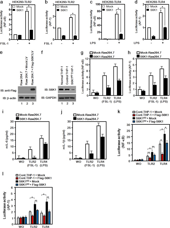
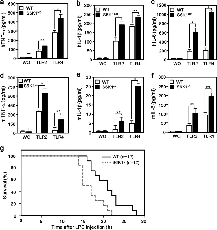
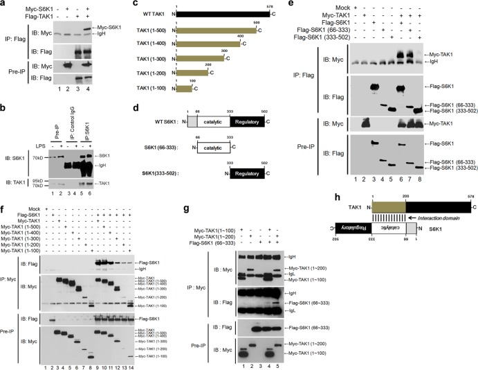
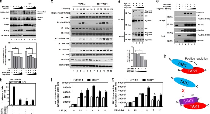
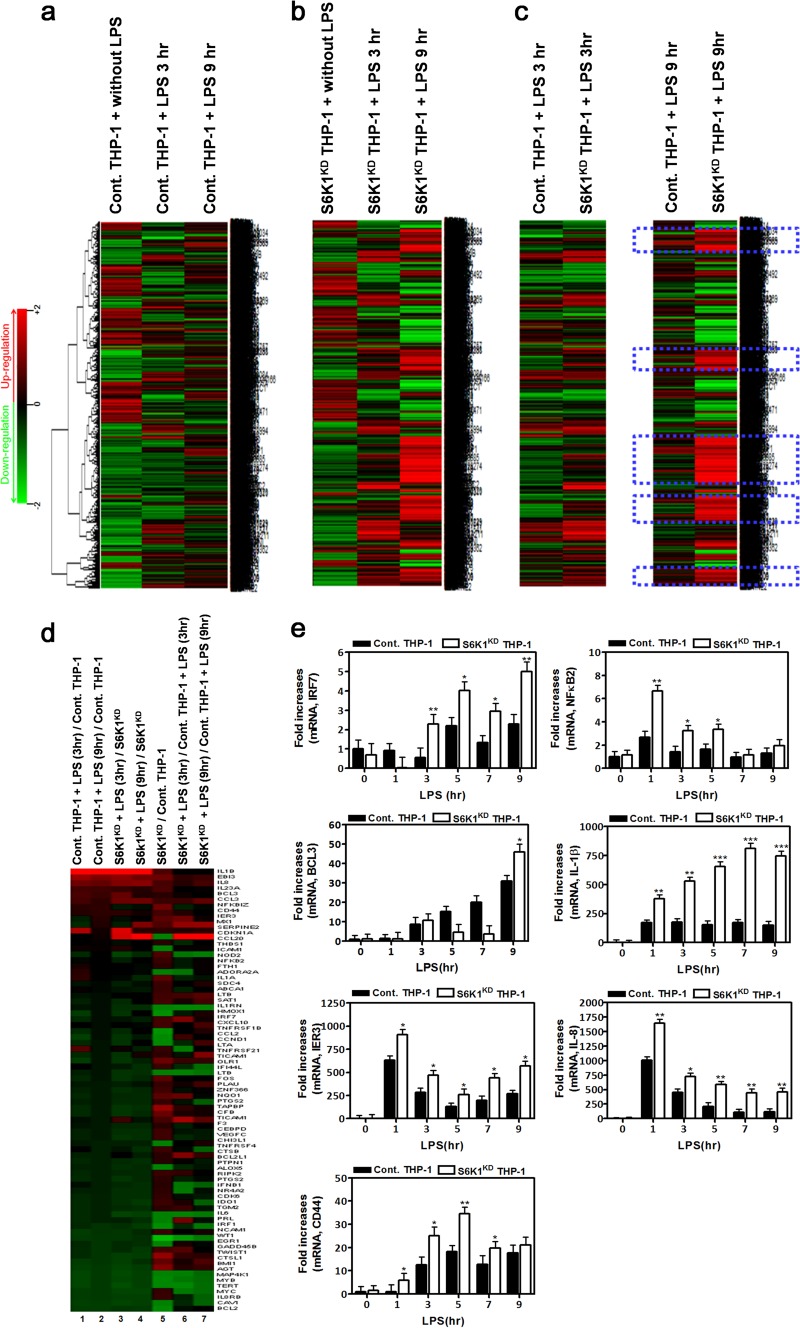
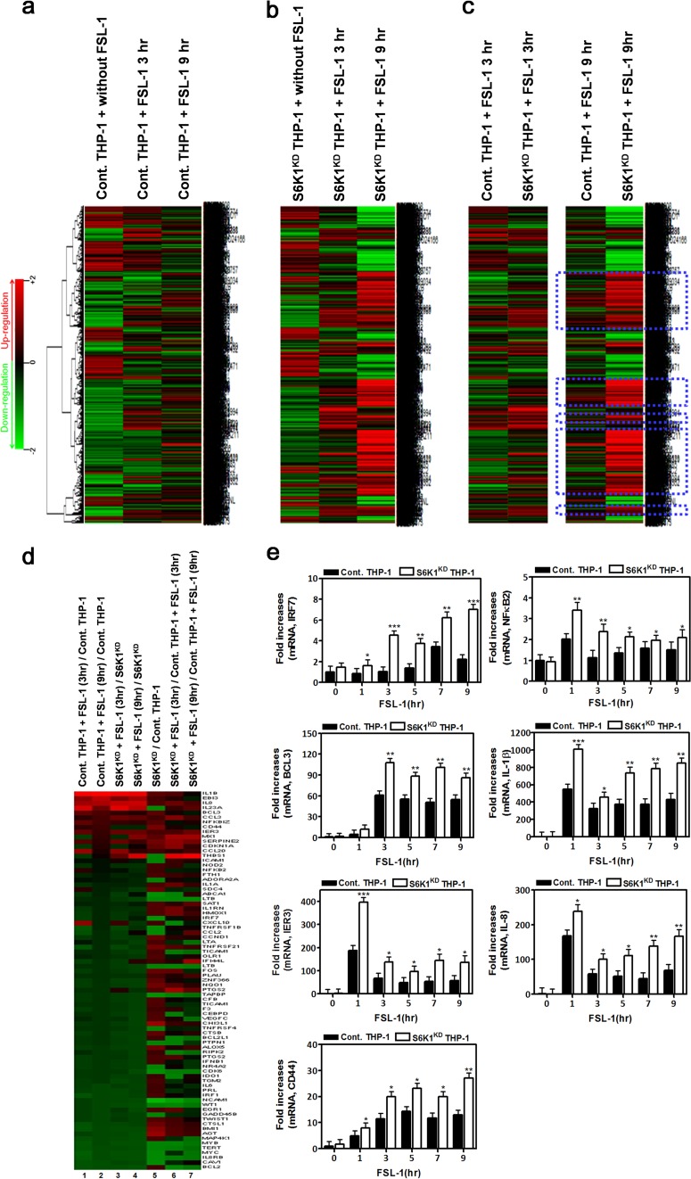
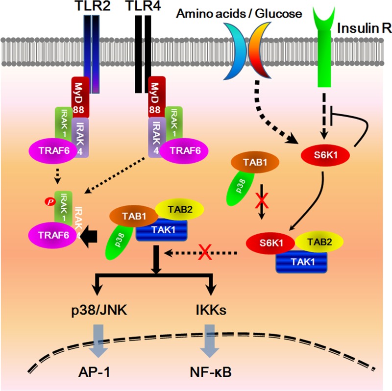
Transforming growth factor β (TGF-β)-activated kinase 1 (TAK1) is a key regulator in the signals transduced by proinflammatory cytokines and Toll-like receptors (TLRs). The regulatory mechanism of TAK1 in response to various tissue types and stimuli remains incompletely understood. Here, we show that ribosomal S6 kinase 1 (S6K1) negatively regulates TLR-mediated signals by inhibiting TAK1 activity. S6K1 overexpression causes a marked reduction in NF-κB and AP-1 activity induced by stimulation of TLR2 or TLR4. In contrast, S6K1(-/-) and S6K1 knockdown cells display enhanced production of inflammatory cytokines. Moreover, S6K1(-/-) mice exhibit decreased survival in response to challenge with lipopolysaccharide (LPS). We found that S6K1 inhibits TAK1 kinase activity by interfering with the interaction between TAK1 and TAB1, which is a key regulator protein for TAK1 catalytic function. Upon stimulation with TLR ligands, S6K1 deficiency causes a marked increase in TAK1 kinase activity that in turn induces a substantial enhancement of NF-κB-dependent gene expression, indicating that S6K1 is negatively involved in the TLR signaling pathway by the inhibition of TAK1 activity. Our findings contribute to understanding the molecular pathogenesis of the impaired immune responses seen in type 2 diabetes, where S6K1 plays a key role both in driving insulin resistance and modulating TLR signaling.


Figures

FIG 1
FIG 1
S6K1 is negatively involved in the activation of NF-κB and AP-1 induced by TLR2 and TLR4. (a to d) HEK293-TLR2 cells (a and b) and HEK293-TLR4 cells (c and d) were cotransfected with a mock or Flag-S6K1 vector together with pBIIx-luc (a and c) or with AP-1-luc together with the Renilla luciferase vector (b and d). Twenty-four hours after transfection, cells were treated with or without FSL-1 (a and b) or LPS (c and d) for 6 h and then analyzed for luciferase activity. Results are expressed as the fold induction in luciferase activity relative to that in untreated cells. (e) The mock or Flag-S6K1 lentivirus vector was used to infect Raw 264.7 cells as described in Materials and Methods. The expression of Flag-S6K1 was examined with anti-Flag antibody by using Western blot assay. Immunoblotting (IB) with anti-β-actin antibody was performed to generate a control for gel loading. (f) A total of 2 × 10⁵ THP-1 cells were cultured in a 24-well plate and infected with lentiviral particles containing shRNA targeting human S6K1 (S6K1KD THP-1) or control shRNA lentiviral particles (Control THP-1). Cells were cultured in medium containing puromycin (4 to 8 μg/ml) for 2 weeks to select stable clones. The knockdown efficacy was examined with anti-S6K1 antibody using Western blotting. Immunoblotting with anti-GAPDH antibody was performed to generate a control for gel loading. (g and h) Mock-Raw 264.7 and S6K1-Raw 264.7 cells were cotransfected with pBIIx-luc (g) or AP1-luc (h) reporter together with Renilla luciferase vector. Twenty-four hours after transfection, cells were treated with or without (WO) FSL-1 or LPS for 6 h and then analyzed for luciferase activity. Results are expressed as the fold induction in luciferase activity relative to that in untreated cells. The data shown are the averages of a minimum of three independent experiments, with error bars denoting standard deviations (±SD). (i and j) Mock-Raw 264.7 and S6K1-Raw 264.7 cells were treated with or without TLR2 or TLR4 agonist (FSL-1 and LPS, respectively) for 9 h, and then supernatants were harvested. Levels of mouse IL-6 and mouse IL-1β were measured in the supernatants according to the assay manufacturer's protocol. (k and l) Control THP-1 and S6K1KD THP-1 cells were transfected with the mock or Flag-S6K1 vector with pBIIx-luc (k) or AP1-luc (l) together with Renilla luciferase vector. Twenty-four hours after transfection, cells were treated with or without FSL-1 or LPS for 6 h and then analyzed for luciferase activity. Results are expressed as the fold induction in luciferase activity relative to that in untreated cells. The data shown are the averages of a minimum of three independent experiments, with error bars denoting ±SD. *P < 0.01, **P < 0.05.
FIG 2
FIG 2
S6K1 deficiency increases proinflammatory cytokine production induced by TLR2 and TLR4. (a to c) Wild-type and S6K1KD THP-1 cells were treated with or without (WO) FLS-1 or LPS for 9 h. Human TNF-α (hTNF-α) (a), hIL-1β (b), and hIL-6 (c) production was analyzed by enzyme-linked immunosorbent assay (ELISA). The data shown are the averages of a minimum of three independent experiments, with error bars denoting ±SD. *, P < 0.01; **, P < 0.05. (d to f) Splenocytes were isolated from wt or S6K1−/− mice (n = 3 each). Cells were plated into 96-well plates (5 × 10⁵ cells/well) and treated with or without TLR2 and TLR4 agonists (FSL-1 and LPS, respectively) for 24 h. Mouse TNF-α (mTNF-α) (d), mIL-1β (e), and mIL-6 (f) production was analyzed by ELISA. Data are shown as the mean ± SD. *, P < 0.01; **, P < 0.05. (g) Six- to 8-week-old S6K1−/− mice and wild-type controls were intraperitoneally injected with a lethal dose of LPS (40 mg/kg of body weight) and monitored for mortality. Survival of mice was analyzed using Kaplan-Meier survival curves, and the difference in median survival times of S6K1−/− and wild-type mice was assessed by log rank tests (P = 0.0064; hazard ratio, 2.55; 95% CI of ratio, 1.05 to 6.22).
FIG 3
FIG 3
The TAK1 N terminus interacts with the catalytic domain of S6K1. (a) HEK293 cells were transfected with Myc-S6K1, Flag-TAK1, or Myc-S6K1 and Flag-TAK1 vector. At 36 h after transfection, cells were extracted and immunoprecipitated (IP) with anti-Flag antibody. The interaction was detected by Western blotting with anti-Myc antibody. The presence of Myc-S6K1 and Flag-TAK1 in the pre-IP lysates was verified by Western blotting. (b) HEK293 cells were treated with or without LPS (100 ng/ml) for 45 min. Cells were extracted and immunoprecipitated with anti-S6K1 or control IgG antibody. The endogenous interaction was detected by Western blotting with anti-TAK1 antibody. (c) Five different Myc-tagged truncated mutants of TAK1 were generated from the control TAK1 vector as described in Materials and Methods. (d) Two different Flag-tagged truncated mutants of S6K1 were generated from the control S6K1 vector as described in Materials and Methods. (e) HEK293 cells were transfected with combinations of proteins as indicated above the lanes. At 36 h after transfection, transfected cells were extracted and immunoprecipitated with anti-Flag antibody. The interaction was detected by Western blotting with anti-Myc antibody. The presence of proteins in the pre-IP lysates was verified by Western blotting as indicated to the right. (f) HEK293 cells were transfected with combinations of proteins as indicated above the lanes. At 36 h after transfection, transfected cells were extracted and immunoprecipitated with anti-Myc antibody. The interaction was detected by Western blotting with anti-Flag antibody. The presence of proteins in the pre-IP lysates was verified by Western blotting as indicated to the right. (g) HEK293 cells were transfected with combinations of proteins as indicated above the lanes. At 36 h after transfection, transfected cells were extracted and immunoprecipitated with anti-Myc antibody. The interaction was detected by Western blotting with anti-Flag antibody. The presence of the proteins in the pre-IP lysates was verified by Western blotting. (h) A model of the interaction between the N terminus of TAK1 and catalytic domain of S6K1.
FIG 4
FIG 4
S6K1 competitively interferes with the binding of TAB1 to TAK1, which inhibits TAK1 kinase activity. (a) HEK293 cells were transfected with Myc-TAK1 and different concentrations of Flag-S6K1. At 36 h after transfection, cells were treated with or without LPS (100 ng/ml) for 45 min, extracted, and Western blotting was performed with the antibodies indicated to the left. The band intensity of pho-TAK1 was analyzed with Image J (bottom). Data shown are the averages from a minimum of three independent experiments (±SD). *, P < 0.05. (b) HEK293-TLR4 cells were cotransfected with wt S6K1, DN-S6K1, or CA-S6K1 vector, together with pBIIx-luc reporter and Renilla luciferase vector. Twenty-four hours after transfection, cells were treated with or without (WO) LPS for 6 h and then analyzed for luciferase activity. Results are expressed as the fold induction in luciferase activity relative to that in untreated cells. The data shown are the averages of a minimum of three independent experiments, with error bars denoting ±SD. (c) Wild-type and S6K1KD THP-1 cells were treated with or without LPS for different times, and then Western blotting was performed as described in Materials and Methods. (d) HEK293 cells were transfected with Myc-TAK1, Flag-S6K1, and different concentrations of Flag-TAB1 as indicated. At 36 h after transfection, cells were extracted and immunoprecipitated with anti-Myc antibody. Interaction was detected by Western blotting with anti-Flag antibody. The presence of Myc-TAK1, Flag-S6K1, and Flag-TAB1 in the pre-IP lysates was verified by Western blotting. The band intensity of Flag-S6K1 was analyzed with Image J (bottom). The data shown are the averages of a minimum of three independent experiments (±SD). *, P < 0.05. (e) HEK293 cells were transfected with Myc-TAK1, Flag-TAB1, and different concentrations of Flag-S6K1 (66-333) as indicated. At 36 h after transfection, cells were extracted and immunoprecipitated with anti-Myc antibody. The interaction was detected by Western blotting with anti-Flag antibody. The presence of Myc-TAK1, Flag-TAB1, and Flag-S6K1 (66-333) in the pre-IP lysates was verified by Western blotting. (f and g) Wild type and S6K1KD THP-1 cells were treated with or without LPS (f) or FSL-1 (g) for different times as indicated. The kinase assay for TAK1 was performed using a c-TAK1 kinase assay kit in accordance with the manufacturer's protocol. The data shown are the averages of a minimum of three independent experiments (±SD). *, P < 0.05; **, P < 0.01. (h) A model for the negative regulation of TAK1. The C terminus of TAB1 interacts with the N terminus of TAK1, and the TAB1 association to TAK1 positively regulates TAK1 activity via the recruitment of p38 and induction of catalytic activity (top). In contrast, the interaction between the internal catalytic domain of S6K1 and the N terminus of TAK1 inhibits the TAB1 interaction with TAK1, which results in the inhibition of TAK1 catalytic activity.
FIG 5
FIG 5
S6K1 knockdown THP-1 cells exhibit a marked induction in NF-κB-dependent gene expression in response to stimulation of TLR4. (a) Control THP-1 cells were treated with or without TLR4 agonist LPS (100 ng/ml) for 3 h or 9 h. Microarray analysis was performed as described in Materials and Methods. (b) S6K1KD THP-1 cells were treated with or without LPS for 3 h or 9 h. Microarray analysis was performed as described in Materials and Methods. (c) Comparison of microarray data between control THP-1 and S6K1KD THP-1 cells treated with LPS for 3 h or 9 h. Highly upregulated genes in S6K1KD THP-1 cells treated with LPS are indicated with dashed blue boxes. (d) Microarray analysis of the NF-κB-dependent upregulated and downregulated genes is shown. The experimental conditions are indicated above the columns. (e) Control THP-1 and S6K1KD THP-1 cells were treated with or without LPS (100 ng/ml) for different times as indicated. Total RNAs were isolated from each sample, and quantitative RT-PCR analysis was performed with specific primers targeted to the genes indicated on the y axes. Data represent the averages of data from two independent experiments, each done with triplicates. Error bars represent the means ± SD based on these six samples. *, P < 0.05; **, P < 0.01; ***, P < 0.001.
FIG 6
FIG 6
S6K1 knockdown THP-1 cells exhibit a marked induction in NF-κB-dependent gene expression in response to stimulation of TLR2. (a) Control THP-1 cells were treated with or without FSL-1 (10 μg/ml) for 3 h or 9 h. Microarray analysis was performed as described in Materials and Methods. (b) S6K1KD THP-1 cells were treated with or without FSL-1 for 3 h or 9 h. Microarray analysis was performed as described in Materials and Methods. (c) Comparison of microarray data between control THP-1 and S6K1KD THP-1 cells treated with FSL-1 for 3 h or 9 h. The highly upregulated genes in S6K1KD THP-1 treated with FSL-1 are indicated with dashed blue boxes. (d) Microarray analysis of the NF-κB-dependent upregulated and downregulated genes. The experimental conditions are indicated above the columns. (e) Control THP-1 and S6K1KD THP-1 cells were treated with or without FSL-1 (100 ng/ml) for different times as indicated. Total RNAs were isolated from each sample, and quantitative RT-PCR analysis was performed with specific primers targeted to the genes indicated on the y axes. Data represent the averages of data from two independent experiments, each done with triplicates. Error bars represent the means ± SD based on these six samples. *, P < 0.05; **, P < 0.01; ***, P < 0.001.
FIG 7
FIG 7
A schematic model for how S6K1 inhibits TLR2- or TLR4-mediated signaling pathways. Upon stimulation of TLR2 or TLR4 receptors, MyD88 binds to the cytoplasmic portion of the TLRs through interactions between individual TIR domains. IRAKs, such as IRAK-4 and IRAK-1, and TRAF6 are recruited to the receptor, and then phosphorylated IRAK-1 (by IRAK-4) dissociates from the receptor together with TRAF6. TRAF6 further interacts with TAK1, TAB1, and TAB2. TAB1 also interacts with p38α through its p38α-binding domain. TAB1 plays several roles in the regulation of the TAK1 complex, such as the recruitment of p38α MAPK to the TAK1 complex for the phosphorylation of TAB1 and induction of TAK1 catalytic activity. The activated TAK1 eventually induces the activation of NF-κB and AP-1 transcription factors through activation of the IKK complex and MAP kinases, respectively. In this study, we speculate on the inhibitory role of S6K1 in type 2 diabetes, inducing insulin resistance under conditions of nutrient overload. Under high-nutrient conditions, S6K1 acts as a negative regulator in insulin signaling. Simultaneously, S6K1 interacts with TAK1, which results in the inhibition of the association of TAB1 and p38α to TAK1. The inhibitory effect eventually induces the inhibition of NF-κB and AP-1 activation by the suppression of IKKs and p38/JNK activation, respectively.

References

    1. Ninomiya-Tsuji J, Kishimoto K, Hiyama A, Inoue J, Cao Z, Matsumoto K. 1999. The kinase TAK1 can activate the NIK-I kappaB as well as the MAP kinase cascade in the IL-1 signalling pathway. Nature 398:252–256. 10.1038/18465 - DOI - PubMed
    1. Shibuya H, Yamaguchi K, Shirakabe K, Tonegawa A, Gotoh Y, Ueno N, Irie K, Nishida E, Matsumoto K. 1996. TAB1: an activator of the TAK1 MAPKKK in TGF-beta signal transduction. Science 272:1179–1182. 10.1126/science.272.5265.1179 - DOI - PubMed
    1. Hasegawa M, Fujimoto Y, Lucas PC, Nakano H, Fukase K, Núñez G, Inohara N. 2008. A critical role of RICK/RIP2 polyubiquitination in Nod-induced NF-kappaB activation. EMBO J. 27:373–383. 10.1038/sj.emboj.7601962 - DOI - PMC - PubMed
    1. Wang C, Deng L, Hong M, Akkaraju GR, Inoue J, Chen ZJ. 2001. TAK1 is an ubiquitin-dependent kinase of MKK and IKK. Nature 412:346–351. 10.1038/35085597 - DOI - PubMed
    1. Ishitani T, Takaesu G, Ninomiya-Tsuji J, Shibuya H, Gaynor RB, Matsumoto K. 2003. Role of the TAB2-related protein TAB3 in IL-1 and TNF signaling. EMBO J. 22:6277–6288. 10.1093/emboj/cdg605 - DOI - PMC - PubMed

Publication types

MeSH terms

Substances