Clinical Trial
. 2013;9(11):e1003754.
doi: 10.1371/journal.ppat.1003754. Epub 2013 Nov 21.

The effects of somatic hypermutation on neutralization and binding in the PGT121 family of broadly neutralizing HIV antibodies

Affiliations
Clinical Trial

The effects of somatic hypermutation on neutralization and binding in the PGT121 family of broadly neutralizing HIV antibodies

Devin Sok et al. PLoS Pathog. 2013.

Erratum in

  • PLoS Pathog. 2013 Dec;9(12). doi:10.1371/annotation/f1f8c791-61e9-45c6-a455-92c6dadf9f06. Kleinstein, Stephen H [corrected to Kleinstein, Steven H]

Abstract

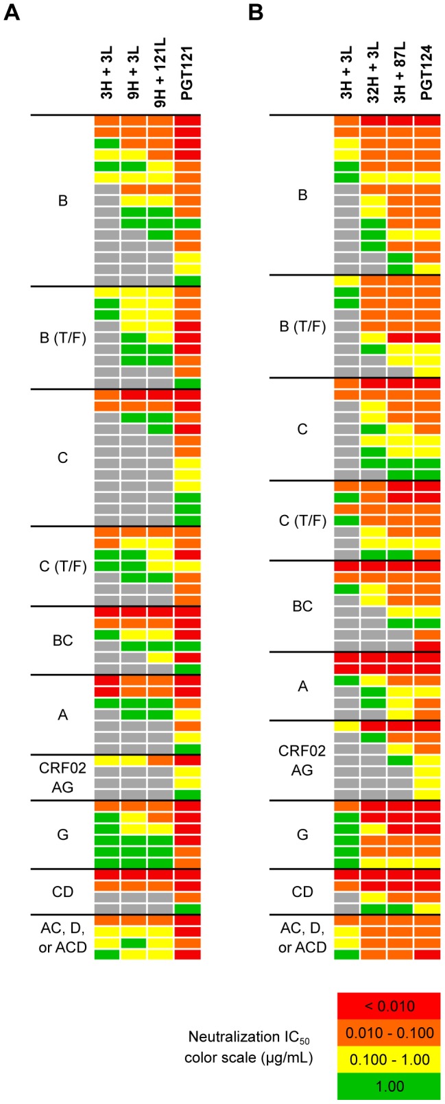
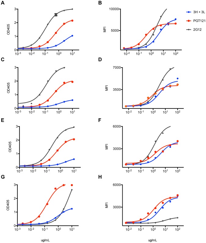
Broadly neutralizing HIV antibodies (bnAbs) are typically highly somatically mutated, raising doubts as to whether they can be elicited by vaccination. We used 454 sequencing and designed a novel phylogenetic method to model lineage evolution of the bnAbs PGT121-134 and found a positive correlation between the level of somatic hypermutation (SHM) and the development of neutralization breadth and potency. Strikingly, putative intermediates were characterized that show approximately half the mutation level of PGT121-134 but were still capable of neutralizing roughly 40-80% of PGT121-134 sensitive viruses in a 74-virus panel at median titers between 15- and 3-fold higher than PGT121-134. Such antibodies with lower levels of SHM may be more amenable to elicitation through vaccination while still providing noteworthy coverage. Binding characterization indicated a preference of inferred intermediates for native Env binding over monomeric gp120, suggesting that the PGT121-134 lineage may have been selected for binding to native Env at some point during maturation. Analysis of glycan-dependent neutralization for inferred intermediates identified additional adjacent glycans that comprise the epitope and suggests changes in glycan dependency or recognition over the course of affinity maturation for this lineage. Finally, patterns of neutralization of inferred bnAb intermediates suggest hypotheses as to how SHM may lead to potent and broad HIV neutralization and provide important clues for immunogen design.


Conflict of interest statement

Authors BBS and EPSJ are employed by 454 Life Sciences - Roche Sequencing Solutions. Authors PYCH and KS are employed by Theraclone-Sciences. FV is employed by, UL and GMC are advisors to, and FV, UL, and GMC own stock in AbVitro, which owns intellectual property in various immune sequencing technologies. The IAVI and Theraclone hold U.S. patent 61/515,528 on the PGT antibodies. All other authors declare no competing interests. This does not alter our adherence to all PLOS Pathogens policies on sharing data and materials.

Figures

Figure 1
Figure 1. Mutation summary of selected heavy and light chain clones.
Mutation frequency was calculated over the V-gene and J-gene as nucleotides or amino acids differing from the putative germline sequence for (A) heavy chain sequences and (B) light chain sequences. The CDR3 regions and insertions and deletions were excluded from the analysis. CDR3 lengths were determined according to the IMGT definition. Analyses were performed with the SciPy stack and figures were generated using matplotlib and graphviz .
Figure 2
Figure 2. PGT121–123 variants identified by deep sequencing were used to build phylogenetic trees using ImmuniTree.
(A) An example identity (y-axis) and mutation (x-axis) plot for PGT121 used to identify PGT121-variants for heavy chain and for (B) light chain; each color represents a unique clone. (C) Heavy chain and (D) light chain SHM phylogenies inferred by the ImmuniTree algorithm. Nodes that were inferred by the algorithm are represented as small circles. Nodes representing observed sequences are depicted as larger circles; node size is proportional to the number of reads assigned to that node. The trees are colored based on level of mutation from germline. Previously isolated affinity matured mAbs are labeled (e.g. PGT122) and nodes selected for synthesis and characterization are labeled blue.
Figure 3
Figure 3. Selected heavy and light chain clones were paired and tested for neutralization breadth and potency on a cross-clade 6-virus panel.
Neutralization breadth and potency summary of all the paired clones. The clones are arranged from least (bottom-left) to most (top-right) mutated with heavy chain listed on the left and light chain listed across the bottom. Boxes are colored according to neutralization score, which is defined as mean(log10(10/IC50)); black boxes represent pairs whose neutralization scores were not determined.
Figure 4
Figure 4. Selected heavy and light chain clones were paired and tested for neutralization breadth and potency on a cross-clade 6-virus panel.
(A) Heavy and light chain nodes leading to mAb PGT121 and (B) PGT124 were paired and tested on a 74-virus panel of PGT121- or PGT124-sensitive viruses. Boxes are colored by IC50 values (µg/ml) of each isolate neutralized.
Figure 5
Figure 5. Higher levels of somatic hypermutation correlates with greater neutralization breadth and potency.
(A) Summary of neutralization data in Figure 4A–B by each clade neutralized. Listed in colored boxes are percentage values. (B) Overall mutation frequencies of mAb combinations, which were calculated by overall number of nucleotide mutations in both heavy and light chains divided by combined heavy and light chain lengths. The mutation frequency of each heavy and light chain was calculated over both V- and J-genes.
Figure 6
Figure 6. Role of somatic hypermutation in neutralization and antibody structure.
(A) Residues from heavy chain intermediates and light chain intermediates were reverted to germline while indel and CDR3 residues were individually mutated to alanines and tested on a cross-clade virus panel. Highlighted in blue are residues that were reverted to germline. Highlighted in orange are residues reverted between intermediates. Filled red dots represent residues reversions that resulted in significant loss of neutralization as single reversions. Empty red dots represent residue reversions that resulted in significant loss of neutralization as pairwise reversions. Reported fold changes in IC50 are shown in Figure S12. (B) Reported fold changes in IC50 for residues reverted to germline in putative intermediate 3H+3L. (C) Residues that were found to be critical for neutralization in (B) were mapped on a putative PGT121 germline crystal structure. Global structural shifts were observed between germline (gray) and PGT121 (colored). Arrows indicate shifts of CDR loops. (D) 11 Å shift of RL94 in the CDRL3, which was identified to be important for neutralization activity. (E) Salt bridge between RH100 in the CDRH3 and DL67 in the FRL3 insertion that contributes to structural shifts. (F) Effect of N51D mutation in CDRL2. NL51 is highly conserved among all antibody variants and substitution completely abrogates neutralization. The introduction of two hydrogen bonds between the side chain of NL51 and backbone atoms might facilitate a helix to loop change in CDRL1. (G) A H34Q substitution in CDRL1 contributes to stabilizing the proximal positions of CDRL1 and CDRL2.
Figure 7
Figure 7. Neutralization activity of PGT122–133 mAbs without the FRL3 insertion and restoring the N-terminus deletion.
The three amino acid insertion in FR3 of the light chain of (A) PGT122, (B) PGT123, (C) PGT124, and (D) PGT133 was removed and paired with the corresponding heavy chains and tested for neutralization activity on a 6-virus panel. Solid lines represent wild-type antibodies with the insertion and dashed lines represent the antibodies without the FR3 insertion. (E) The 7 amino acid N-terminus sequence of IGHV4-59 germline was placed back into the N-terminus of PGT121 and tested for neutralization breadth and potency on a cross-clade 6 virus panel. Solid lines represent WT PGT121 (with N-terminal deletion) and dashed lines represent PGT121 with the N-terminus restored.
Figure 8
Figure 8. Putative germline of PGT121 does not bind monomeric gp120 or cell surface Env.
(A) PGT121germline does not bind recombinant gp120 (92BR020). Recombinant gp120s were produced in 293F cells and purified by lectin column before use in ELISA binding assays. ELISA values are reported in optical density at 405 nm (OD405). (B) PGT121germline does not cell surface Env (92BR020). Cell surface Env was produced by transfecting pseudovirus in 293T cells and binding was measured by flow cytometry (reported in mean fluorescence intensity or MFI).
Figure 9
Figure 9. Inferred intermediate antibodies preferentially bind native Env relative to monomeric gp120.
mAbs 3H+3L (blue) and PGT121 (red) were tested for binding by ELISA to monomeric gp120, which was extracted from lysed virus supernatants: (A) 92BR020, (C) 92RW020, (E) JR-FL E168K/N192A, (G) IAVI C22. Antibodies were also tested for cell surface Env binding by flow cytometry: (B) 92BR020, (D) 92RW020, (F) JR-FL E168K/N192A, (H) IAVI C22. mAb 2G12 was included as a control (gray). ELISA values are reported as optical density at 405 nm (OD405) and flow cytometry values are reported as mean fluorescence intensity (MFI).
Figure 10
Figure 10. Antibody 3H+3L likely crosslinks between trimers.
(A) 3H+3L binds an epitope overlapping with those of PGT121, PGT128, and 2G12 as shown by competition of biotinylated antibody 3H+3L with an antibody panel. Binding assays were performed by flow cytometry on JR-FLΔCT isolate transfected in 293T cells. (B) IgG and Fab fragments were tested for binding on JR-FLΔCT isolate expressed on transfected 293T cells and no substantial differences in avidity were observed. Solid lines represent IgG and dashed lines represent Fab fragments. (C) Purified IgGs of 3H+3L and PGT121 were digested into Fab fragments using Lys-C, purified, and tested for neutralization on a cross-clade panel. Loss of neutralization was found for 3H+3L Fab, but not for PGT121 Fab. Reported values are IC50 ratios of Fab compared to IgG using the equation: (IC50 Fab)∶(IC50 IgG).
Figure 11
Figure 11. Neutralization assays on JR-FL glycan mutants indicate binding of inferred intermediate antibodies to both N301 and N332 on HIV-1 Env.
(A) Neutralization curves of inferred intermediate antibodies on wild-type JR-FL virus (B) JR-FL N301A mutant virus, and (C) JR-FL N332A mutant virus. (D) Neutralization curves of 3H+3L and (E) PGT121 on wild-type JR-FL virus compared to single and double glycan mutant viruses. (F) Neutralization curves of PGT121 on wild-type 92BR020 virus compared to single and double glycan mutant viruses.

References

    1. Kwong PD, Mascola JR (2012) Human antibodies that neutralize HIV-1: identification, structures, and B cell ontogenies. Immunity 37: 412–425 doi:10.1016/j.immuni.2012.08.012 - DOI - PMC - PubMed
    1. Walker LM, Phogat SK, Chan-Hui P-Y, Wagner D, Phung P, et al. (2009) Broad and potent neutralizing antibodies from an African donor reveal a new HIV-1 vaccine target. Science 326: 285–289 doi:10.1126/science.1178746 - DOI - PMC - PubMed
    1. Wu X, Yang Z-Y, Li Y, Hogerkorp C-M, Schief WR, et al. (2010) Rational design of envelope identifies broadly neutralizing human monoclonal antibodies to HIV-1. Science 329: 856–861. - PMC - PubMed
    1. Zhou T, Georgiev I, Wu X, Yang Z-Y, Dai K, et al. (2010) Structural basis for broad and potent neutralization of HIV-1 by antibody VRC01. Science 329: 811–817 doi:10.1126/science.1192819 - DOI - PMC - PubMed
    1. Walker LM, Huber M, Doores KJ, Falkowska E, Pejchal R, et al. (2011) Broad neutralization coverage of HIV by multiple highly potent antibodies. Nature 477: 466–470. - PMC - PubMed

Publication types

Substances