. 2013;9(11):e1003773.
doi: 10.1371/journal.ppat.1003773. Epub 2013 Nov 21.

Type I and type III interferons drive redundant amplification loops to induce a transcriptional signature in influenza-infected airway epithelia

Affiliations

Type I and type III interferons drive redundant amplification loops to induce a transcriptional signature in influenza-infected airway epithelia

Stefania Crotta et al. PLoS Pathog. 2013.

Abstract

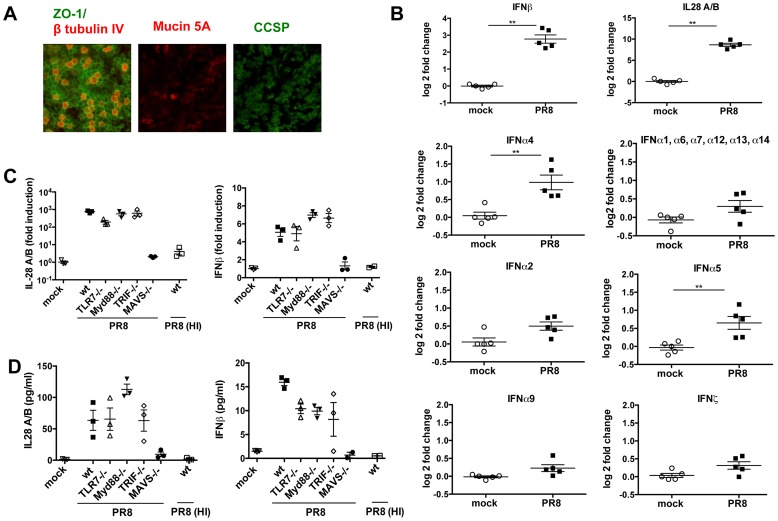
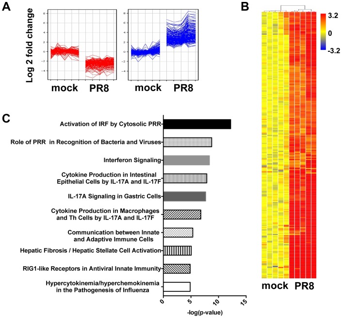
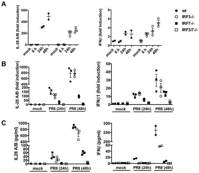
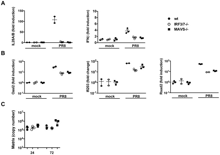
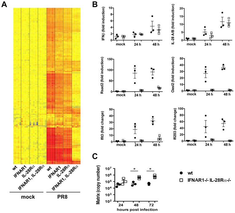
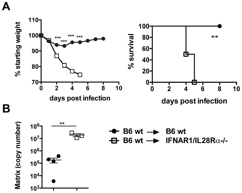
Interferons (IFNs) are a group of cytokines with a well-established antiviral function. They can be induced by viral infection, are secreted and bind to specific receptors on the same or neighbouring cells to activate the expression of hundreds of IFN stimulated genes (ISGs) with antiviral function. Type I IFN has been known for more than half a century. However, more recently, type III IFN (IFNλ, IL-28/29) was shown to play a similar role and to be particularly important at epithelial surfaces. Here we show that airway epithelia, the primary target of influenza A virus, produce both IFN I and III upon infection, and that induction of both depends on the RIG-I/MAVS pathway. While IRF3 is generally regarded as the transcription factor required for initiation of IFN transcription and the so-called "priming loop", we find that IRF3 deficiency has little impact on IFN expression. In contrast, lack of IRF7 reduced IFN production significantly, and only IRF3(-/-)IRF7(-/-) double deficiency completely abolished it. The transcriptional response to influenza infection was largely dependent on IFNs, as it was reduced to a few upregulated genes in epithelia lacking receptors for both type I and III IFN (IFNAR1(-/-)IL-28Rα(-/-)). Wild-type epithelia and epithelia deficient in either the type I IFN receptor or the type III IFN receptor exhibit similar transcriptional profiles in response to virus, indicating that none of the induced genes depends selectively on only one IFN system. In chimeric mice, the lack of both IFN I and III signalling in the stromal compartment alone significantly increased the susceptibility to influenza infection. In conclusion, virus infection of airway epithelia induces, via a RIG-I/MAVS/IRF7 dependent pathway, both type I and III IFNs which drive two completely overlapping and redundant amplification loops to upregulate ISGs and protect from influenza infection.


Conflict of interest statement

The authors have declared that no competing interests exist.

Figures

Figure 1
Figure 1. In primary MTEC, type I and III IFNs are induced upon influenza virus A infection, in a MAVS and replication dependent way.
(A) Mouse tracheal epithelial cells were costained for ZO-1 (green) and β tubulin IV (red), or Mucin 5A (red) and Clara cell secretory protein (CCSP) (green). Images were acquired at ×20 magnification. (B) Total RNA from mock infected and PR8 infected (moi = 0.3) MTEC was analysed using Affymetrix Mouse Genome 430 2.0 microarrays. The signal intensity of each probe was first normalized on the median intensity of that probe across the control group and then represented as log2 fold change relative to the controls. Asterisks indicated statistically significant differences (unpaired t test; **, p<0.01). (C) qRT-PCR analysis of IL-28A/B and IFNβ1 transcripts of MTEC derived from wild-type, TLR7−/−, MyD88−/−, TRIF−/− and MAVS−/− mice, mock infected or infected with either PR8 or heat inactivated PR8. Fold induction is relative to mock treated samples at 24 hpi +/− SEM. (D) IL-28A/B level in the supernatants of the indicated cultures were measured by ELISA 24 hpi.
Figure 2
Figure 2. In primary MTEC, influenza A infection induces an IFN signature.
Total RNA from mock and PR8 infected cells was analysed using Affymetrix Mouse Genome 430 2.0 microarrays at 24(≥4-fold change relative to mock infected; t-test unpaired, p<0.01, Benjamini-Hochberg multiple test correction). (A) K-means clustering of the differentially expressed genes. (B) Heat map of the 234 upregulated-gene, as shown in (A). The range of fold changes is expressed in a log2 scale. (C) Ingenuity Pathway Analysis of the upregulated genes in influenza infected MTEC.
Figure 3
Figure 3. IRF3 is not required for IFN induction in primary MTEC.
(A) MTEC derived from wild-type or IRF3−/− mice were mock treated or infected with PR8. RNA was analyzed at the indicated time by qRT-PCR, normalized to the amount of HPRT transcripts and measured as fold induction relative to the level in the corresponding control samples. (B) Expression of IL-28A/B and IFNβ was measured by qRT-PCR at 24 hpi, in epithelial cultures of the indicated genotypes. (C) Protein levels of IL-28A/B in the supernatants of mock and PR8 infected cells were measured by ELISA 24 hpi or, where indicated, at 48 hpi. Error bars indicate the SEM of replicates.
Figure 4
Figure 4. ISGs are induced in the absence of detectable type I and III IFNs.
Wild-type, MAVS−/− and IRF3−/−IRF7−/− MTEC were either mock infected or infected with PR8 and their RNA analyzed 24 hpi for the induction of (A) both IFNs (IL-28A/B and IFNβ) and (B) ISGs (Oasl2, Ifi203, Rsad2). All the transcripts were first normalized to HPRT levels and then expressed as fold induction relative to the mean of mock infected controls, +/− SEM. (C) Viral replication was measured by qRT-PCR on the Influenza A Matrix gene and expressed as copy number +/− SEM.
Figure 5
Figure 5. Type I and III are redundant in the MTEC system.
(A) Total RNA from mock and PR8 infected cells was analysed using Affymetrix Mouse Genome 430 2.0 microarrays at 24 hpi. Supervised analysis was performed using statistical filtering (≥4-fold change relative to mock infected wild-type in at least one treatment group; 2-way ANOVA, p<0.01, Benjamini-Hochberg multiple test correction). (A) Heat map of the upregulated genes. (B) Quantitative RT-PCR analysis of RNA samples extracted from mock and PR8 infected wild-type and IFNAR1−/−IL-28Rα−/− double knock-out MTEC at 24 and 48 hpi. All transcripts were first normalized to HPRT levels and then expressed as fold induction relative to the mean of mock infected controls, +/− SEM. (C) Wild-type and IFNAR1−/−IL-28Rα−/− double knock-out MTEC were infected and RNA samples collected at the indicated time points. Viral replication was measured by qRT-PCR on the Influenza A Matrix gene and expressed as copy number +/− SEM. Asterisks indicate differences that are statistically significant (unpaired t test; *, P<0.05).
Figure 6
Figure 6. Lack of type I and III IFN signalling in the stromal compartment increases susceptibility to IAV infection.
Chimeric mice were infected i.n. with 105 TCID50 PR8, (A) Weight loss and mortality were measured. Graphs show mean ± SEM and are representative of 2 independent experiments (n = 6). (B) Viral replication was assessed by qRT-PCR on total lung RNA at 4 days post infection. *** p<0.001, ** p<0.01 by 2-way ANOVA with Bonferroni post tests (weight loss), Log-rank (Mantel-Cox) Test (survival) or unpaired t test (RT-PCR quantification).
Figure 7
Figure 7. Basal expression of signalling intermediates is similar in wild-type and IFNAR1−/−IL-28Rα−/− double knock-out MTEC.
(A) Relative expression of IRF3, IRF1, IRF7 and IRF9 was determined in both mock infected and PR8 infected MTEC by qRT-PCR. The transcripts quantities are expressed as ratio to HPRT levels +/− SEM. (B) The same RNA samples were analyzed for STAT1, STAT2, STAT3, STAT4 and STAT6 expression.
Figure 8
Figure 8. Residual IFN production is responsible for ISG induction in MAVS−/− epithelia.
(A) qRT-PCR analysis of Rsad2, Oasl2 and STAT1 in MAVS−/− epithelia infected with PR8 in the presence or absence of blocking anti-IFNAR and neutralizing anti-IL28A/B antibodies. (B) Wild-type epithelia were PR8-infected in the presence or absence of brefeldin A (2.5 µg/ml). Expression of the indicated genes was determined by qRT-PCR at 24 hpi. All transcripts were normalized to HPRT levels and then expressed as fold induction relative to the mean of mock infected controls, +/− SEM. Asterisks indicate differences that are statistically significant (unpaired t test; *, P<0.05; **, P<0.01).

References

    1. Isaacs A, Lindenmann J (1957) Virus interference. I. The interferon. Proc R Soc Lond B Biol Sci 147: 258–267. - PubMed
    1. Kato H, Sato S, Yoneyama M, Yamamoto M, Uematsu S, et al. (2005) Cell type-specific involvement of RIG-I in antiviral response. Immunity 23: 19–28. - PubMed
    1. Rehwinkel J, Tan CP, Goubau D, Schulz O, Pichlmair A, et al. (2010) RIG-I detects viral genomic RNA during negative-strand RNA virus infection. Cell 140: 397–408. - PubMed
    1. Kawai T, Takahashi K, Sato S, Coban C, Kumar H, et al. (2005) IPS-1, an adaptor triggering RIG-I- and Mda5-mediated type I interferon induction. Nat Immunol 6: 981–988. - PubMed
    1. Meylan E, Curran J, Hofmann K, Moradpour D, Binder M, et al. (2005) Cardif is an adaptor protein in the RIG-I antiviral pathway and is targeted by hepatitis C virus. Nature 437: 1167–1172. - PubMed

Publication types

MeSH terms