. 2013;9(11):e1003775.
doi: 10.1371/journal.ppat.1003775. Epub 2013 Nov 21.

SPOC1-mediated antiviral host cell response is antagonized early in human adenovirus type 5 infection

Affiliations

SPOC1-mediated antiviral host cell response is antagonized early in human adenovirus type 5 infection

Sabrina Schreiner et al. PLoS Pathog. 2013.

Abstract

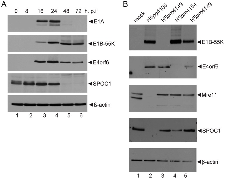
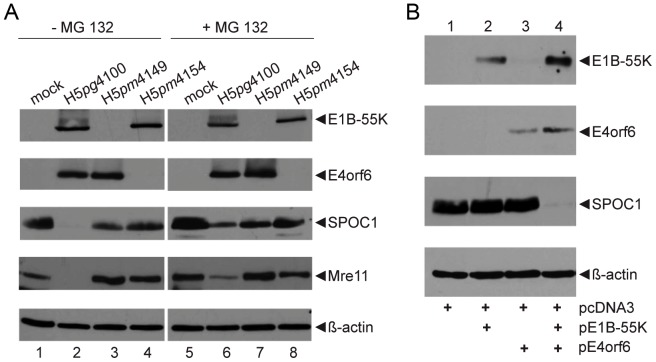
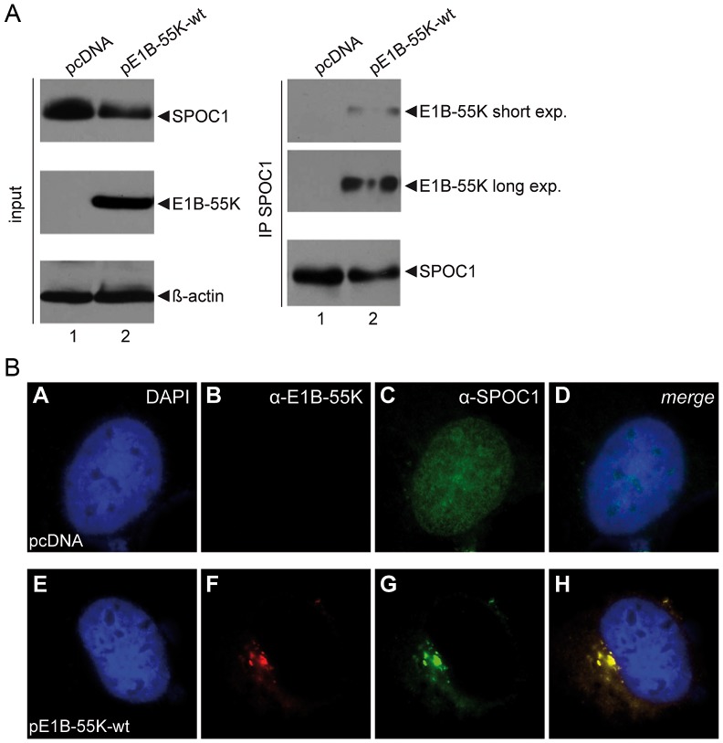
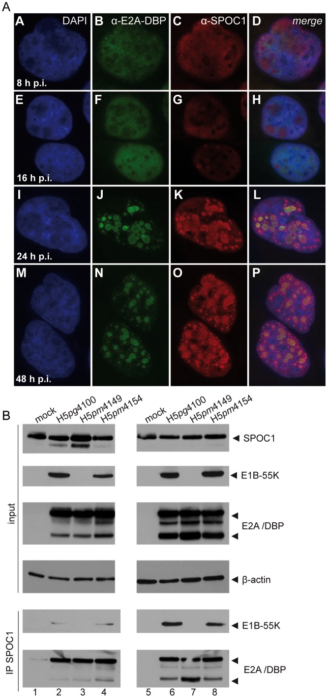
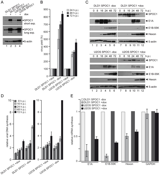
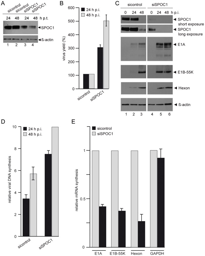
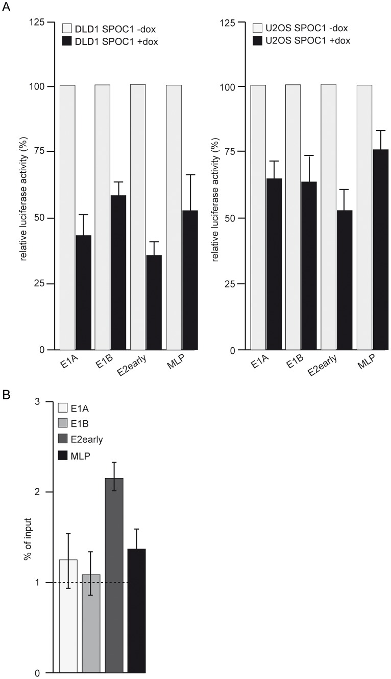
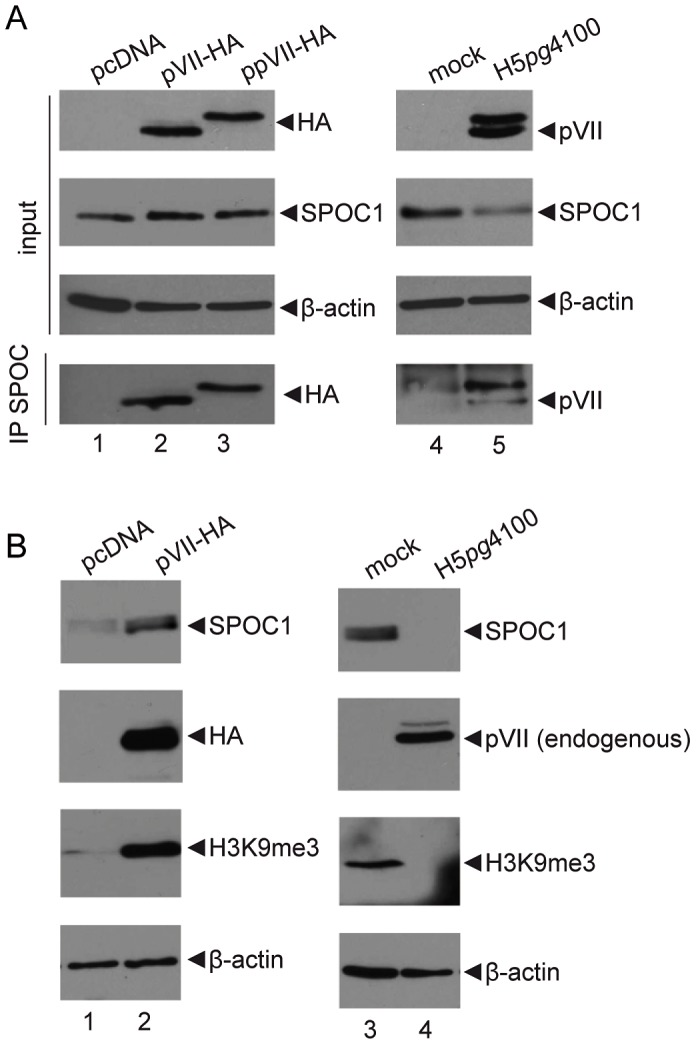
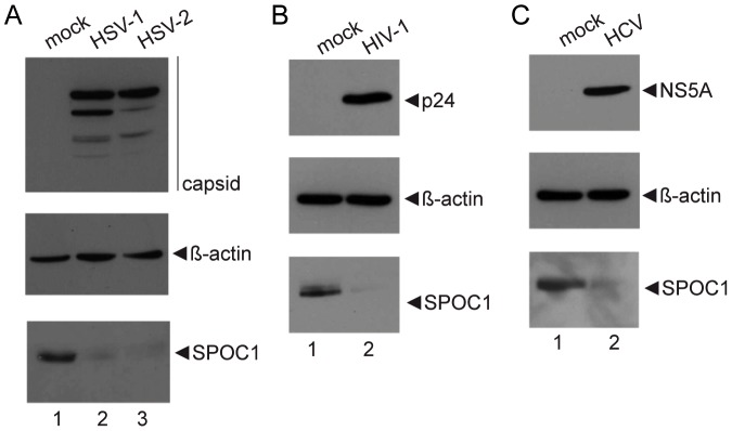
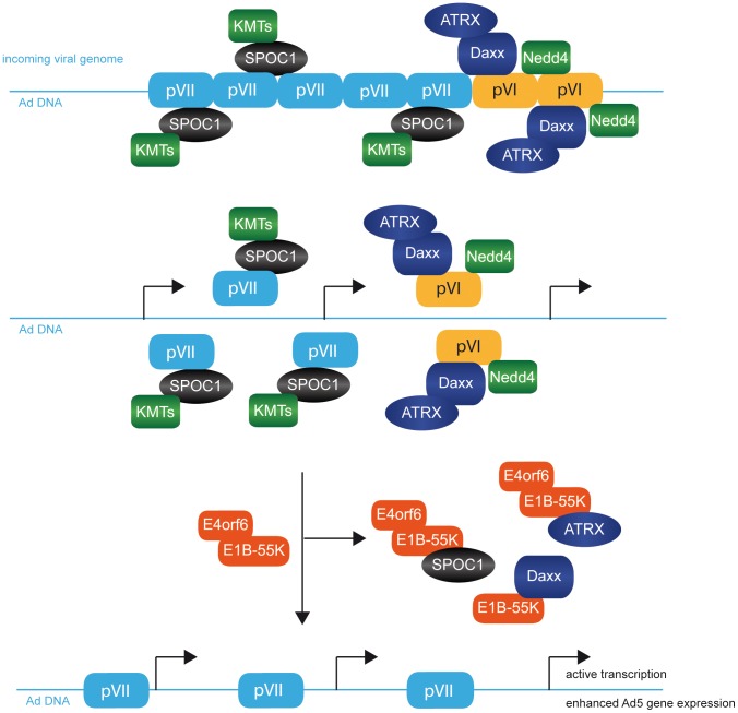
Little is known about immediate phases after viral infection and how an incoming viral genome complex counteracts host cell defenses, before the start of viral gene expression. Adenovirus (Ad) serves as an ideal model, since entry and onset of gene expression are rapid and highly efficient, and mechanisms used 24-48 hours post infection to counteract host antiviral and DNA repair factors (e.g. p53, Mre11, Daxx) are well studied. Here, we identify an even earlier host cell target for Ad, the chromatin-associated factor and epigenetic reader, SPOC1, recently found recruited to double strand breaks, and playing a role in DNA damage response. SPOC1 co-localized with viral replication centers in the host cell nucleus, interacted with Ad DNA, and repressed viral gene expression at the transcriptional level. We discovered that this SPOC1-mediated restriction imposed upon Ad growth is relieved by its functional association with the Ad major core protein pVII that enters with the viral genome, followed by E1B-55K/E4orf6-dependent proteasomal degradation of SPOC1. Mimicking removal of SPOC1 in the cell, knock down of this cellular restriction factor using RNAi techniques resulted in significantly increased Ad replication, including enhanced viral gene expression. However, depletion of SPOC1 also reduced the efficiency of E1B-55K transcriptional repression of cellular promoters, with possible implications for viral transformation. Intriguingly, not exclusive to Ad infection, other human pathogenic viruses (HSV-1, HSV-2, HIV-1, and HCV) also depleted SPOC1 in infected cells. Our findings provide a general model for how pathogenic human viruses antagonize intrinsic SPOC1-mediated antiviral responses in their host cells. A better understanding of viral entry and early restrictive functions in host cells should provide new perspectives for developing antiviral agents and therapies. Conversely, for Ad vectors used in gene therapy, counteracting mechanisms eradicating incoming viral DNA would increase Ad vector efficacy and safety for the patient.


Conflict of interest statement

The authors have declared that no competing interests exist.

Figures

Figure 1
Figure 1. SPOC1 is reduced during Ad infection.
H1299 cells were infected with wild type H5pg4100 at a multiplicity of 50 FFU per cell. Cells were harvested after indicated time points post infection, total-cell extracts were prepared, separated by SDS-PAGE and subjected to immunoblotting. (A) Immunoblotting using mouse monoclonal antibody M73 (E1A), 2A6 (E1B-55K), RSA3 (E4orf6), rat monclonal SPOC1 antibody and mouse monoclonal antibody AC-15 (β-actin) as a loading control. (B) H1299 cells were infected with wildtype (H5pg4100) and mutant viruses (H5pm4149, H5pm4154, H5pm4139) at moi of 50 FFU per cell. Cells were harvested after 48 hours, and immunoblotted using the antibodies above, plus rabbit polyclonal Mre11 antibody.
Figure 2
Figure 2. Proteasomal degradation of SPOC1 by the E1B-55K/E4orf6 E3 ubiquitin ligase complex.
(A) H1299 cells were infected with wildtype (H5pg4100) and mutant viruses (H5pm4149, H5pm4154) at moi of 50 FFU per cell. Cells were treated for six hours with proteasome inhibitor (+MG 132), before total-cell extracts were prepared 48 h p.i.. Proteins were separated by SDS-PAGE and subjected to immunoblotting using mouse monoclonal antibody 2A6 (E1B-55K), RSA3 (E4orf6), rat monclonal SPOC1 antibody and mouse monoclonal antibody AC-15 (β-actin) as a loading control. (B) H1299 cells were transfected with pcDNA3-derived plasmids expressing wildtype E1B-55K, E4orf6, or a combination of both. Cells were harvested 48 hours post infection. Total cell extracts were prepared and specific proteins were immunoblotted as described in A.
Figure 3
Figure 3. SPOC1 is a novel interaction partner of viral E1B-55K protein.
(A) H1299 cells were transfected with empty control plasmid or wild type E1B-55K (pE1B-55K-wt). 24 hours post transfection total cell extracts were prepared. E1B-55K was immunoprecipitated using rabbit polyclonal SPOC1 ab. Proteins were separated on 10% SDS-PAGE and visualized by immunoblotting. Input levels of total cell lysates and co-precipitated proteins were detected using monoclonal antibody 2A6 (E1B-55K), SPOC1-specific rat monoclonal antibody and mouse monoclonal antibody AC-15 (β-actin) as a loading control. (B) H1299 cells were transfected with plasmid constructs encoding wild type E1B-55K. After methanol fixation, the cells were labeled with mouse monoclonal antibody 2A6 (E1B-55K) and SPOC1 specific rat monoclonal antibody. Primary antibodies were detected with Texas Red and FITC conjugated secondary antibody. Representative staining patterns are shown. Nuclei are visualized by DAPI staining (magnification ×7600).
Figure 4
Figure 4. SPOC1 is recruited to Ad replication centers during infection.
(A) Human DLD1 cells, which stably overexpress SPOC1 after doxycyclin treatment to overcome degradation of the cellular factor, were infected with wildtype H5pg4100 at a moi of 10 FFU per cell; throughout the experiment the cells were grown in media supplied with doxycyclin. After indicated time points, cells were fixed with methanol and labeled with E2A/DBP mouse mab B6-8 and SPOC1-specific rat monoclonal antibody. Primary antibodies were detected with Texas Red or FITC conjugated secondary antibody. Representative staining patterns are shown. Nuclei are visualized by DAPI staining (magnification ×7600). Additionally, merge panels show colocalization of the indicated proteins. (B) DLD1 (left panel) and U2OS (right panel) cells, which stably overexpress SPOC1 after doxycyclin treatment were infected with wildtype (H5pg4100) and mutant viruses (H5pm4149, H5pm4154) at moi of 50 FFU per cell. Cells were treated with doxycyclin 12 hours before infection and throughout the experiment to stably induce SPOC1 expression, before total cell extracts were prepared 24 hours post infection. E1B-55K and E2A/DBP were immunoprecipitated using rabbit polyclonal SPOC1 antibody. Proteins were separated on 10% SDS-PAGE and visualized by immunoblotting. Input levels of total-cell lysates and co-precipitated proteins were detected using monoclonal antibody 2A6 (E1B-55K), B6-8 (E2A/DBP), SPOC-1-specific rat monoclonal antibody, and mouse monoclonal antibody AC-15 (β-actin) as a loading control.
Figure 5
Figure 5. SPOC1 overexpression represses Ad5 gene expression.
(A) To validate the model system, DLD1 or U2OS cells were treated with doxycyclin 12 hours before infection to induce SPOC1 expression; throughout the experiment the cells were grown in media supplied with doxycyclin. Cell extracts were subjected to 10% PAGE and immunoblotting using rat monclonal SPOC1 antibody and mouse monoclonal antibody AC-15 (β-actin) as a loading control. (B) DLD1/U2OS cells were infected with wild type H5pg4100 at a multiplicity of 50 FFU per cell. Viral particles were harvested at the indicated time-points after infection and virus yield was determined by quantitative E2A-72K immunofluorescence staining on HEK293 cells. The results represent the average from three independent experiments and were normalized to values for particle synthesis in SPOC1 induced cells infected with wild type H5pg4100. (C) DLD1/U2OS cells were infected with wild type H5pg4100 at a multiplicity of 50 FFU per cell. Total-cell extracts were prepared at the indicated times post infection. Proteins were separated by 10% SDS-PAGE, and immunoblotted with mouse monoclonal antibody M73 (E1A), 2A6 (E1B-55K), anti-Ad5 rabbit polyclonal serum L133, rat monclonal SPOC1 antibody and mouse monoclonal antibody AC-15 (β-actin) as a loading control. (D) DLD1/U2OS cells were infected with wild type H5pg4100 at a multiplicity of 50 FFU per cell. Total cell extracts were prepared and treated with proteinase K. PCR was performed using E1B-specific primers (E1B-fw 3′-CGC GGG ATC CAT GGA GCG AAG AAA CCC ATC TGA GC-5′; E1B-rev 3′-CGG TGT CTG GTC ATT AAG CTA AAA-5′). The same amounts of PCR product were separated on an analytic agarose gels (1%) and quantification was achieved with the Gene Snap Software (Syngene). The results shown represent the averages from three independent experiments. (E) DLD1/U2OS cells were infected with wild type H5pg4100 at a multiplicity of 50 FFU per cell. 24 h p.i. total RNA was extracted, reverse transcribed and quantified by RT-PCR using primers specific for E1A (E1A fwd: 5′ GTGCCCCATTAAACCAGTTG 3′; E1A rev: 5′ GGCGTTTACAGCTCAAGTCC 3′), E1B-55K (E1B fwd: 5′-GAGGGTAACTCCAGGG TGCG-3′; E1B rev: 5′-TTTCACTAGCATGAAGCAACCACA-3′), hexon (hexon rev: 5′-GAACGGTGTGCGCAGGTA-3′; hexon fwd 5′-CGCTGGACATGACTTTTG AG-3′) and GAPDH (GAPDH fwd:5′-ACCACAGTCCATGCCATCAC-3′ rev:5′-TCCACCACCCTGTTGCTGTA-3′). Data were normalized to 18S rRNA levels (18S rRNA fwd: 5′-CGGCTACCACATCCAAGGAA-3′; 18S rRNA rev: 5′-GCTGGAATTACCGCGGCT-3′). Values correspond to the mean of triplicates and error bars indicate the standard error of the mean.
Figure 6
Figure 6. SPOC1 depletion enhances Ad5 gene expression.
(A) H1299 cells were transfected with siRNA constructs against SPOC1 (si768, 5′-UCA CCU GUC CUG UGC GAA AdTdT-3′) and control siRNA (5′-AGG UAG UGU AAU CGC CUU GdTdT-3′). Cells were harvested at 24 and 48 hours post transfection. Total cell extracts were prepared and proteins were separated by SDS-PAGE and subjected to immunoblotting using SPOC1-specific rat monoclonal ab. β-actin was included as a loading control. (B) H1299 cells were treated with indicated siRNAs to induce SPOC1 depletion and infected 24 hours post transfection with wild type H5pg4100 at a multiplicity of 50 FFU per cell. Viral particles were harvested at the indicated time-points after infection and virus yield was determined by quantitative E2A-72K immunofluorescence staining on HEK293 cells. The results represent the average from three independent experiments. (C) H1299 cells were infected with wild type H5pg4100 at a multiplicity of 50 FFU per cell. Total-cell extracts were prepared at the indicated times post infection. Proteins were separated by 10% SDS-PAGE, and immunoblotted with mouse monoclonal antibody M73 (E1A), 2A6 (E1B-55K), anti-Ad5 rabbit polyclonal serum L133, rat monclonal SPOC1 antibody and mouse monoclonal antibody AC-15 (β-actin) as a loading control. (D) H1299cells were infected with wild type H5pg4100 at a multiplicity of 50 FFU per cell. Total cell extracts were prepared and treated with proteinase K. PCR was performed using E1B-specific primers (E1B-fw 3′-CGC GGG ATC CAT GGA GCG AAG AAA CCC ATC TGA GC-5′; E1B-rev 3′-CGG TGT CTG GTC ATT AAG CTA AAA-5′). The same amounts of PCR product were separated on an analytic agarose gels (1%) and quantification was achieved with the Gene Snap Software (Syngene). The results shown represent the averages from three independent experiments. (E) H1299 cells were infected with wild type H5pg4100 at a multiplicity of 50 FFU per cell. 24 h p.i. total RNA was extracted, reverse transcribed and quantified by RT-PCR using primers specific for E1A (E1A fwd: 5′ GTGCCCCATTAAACCAGTTG 3′; E1A rev: 5′ GGCGTTTACAGCTCAAGTCC 3′), E1B-55K (E1B fwd: 5′-GAGGGTAACTCCAGGG TGCG-3′; E1B rev: 5′-TTTCACTAGCATGAAGCAACCACA-3′), hexon (hexon rev: 5′-GAACGGTGTGCGCAGGTA-3′; hexon fwd 5′-CGCTGGACATGACTTTTGAG-3′) and GAPDH (GAPDH fwd:5′-ACCACAGTCCATGCCATCAC-3′ rev:5′-TCCACCACCCTGTTGCTGTA-3′). Data were normalized to 18S rRNA levels (18S rRNA fwd: 5′-CGGCTACCACATCCAAGGAA-3′; 18S rRNA rev: 5′-GCTGGAATTACCGCGGCT-3′). Values correspond to the mean of triplicates and error bars indicate the standard error of the mean.
Figure 7
Figure 7. SPOC1 represses viral transcription.
DLD1 or U2OS cells were treated with doxycyclin for 12(A) Transfection with luciferase reporter plasmids under Ad promoter control (E1A, E1B, E2early, MLP). 48 h after transfection, samples were lysed and absolute luciferase activity was measured. All samples were normalized for transfection efficiency by measuring Renilla-luciferase activity. Activity of empty promotor constructs, E1A, E1B, E2early and MLP promoters in non-treated and non-overxpressing cells was normalized to 100%. Mean and STD are from three independent experiments. (B) After infection with H5pg4100 Ad5 wildtype at moi of 20 FFU/cell. 24 hours post infection SPOC1-induced cells were fixed with formaldehyde and analyzed by ChIP assays (see Material and Methods). The average Ct-value was determined from triplicate reactions and normalized against non-specific IgG controls with standard curves for each primer pair. The y-axis indicates the percentage of immunoprecipitated signal from the input ( = 100%). The dotted line highlights values above 1% of input, commonly stated as significant chromatin/protein binding.
Figure 8
Figure 8. SPOC1 functionally cooperates with Ad core protein pVII.
(A) H1299 cells were transfected with pVII expression constructs (ppVII: prepVII; pVII: processed pVII) and infected with wildtype (H5pg4100) at moi of 50 FFU per cell. Total cell extracts were prepared 24 hours post transfection and infection. pVII was immunoprecipitated using anti SPOC1 rabbit polyclonal antibody. Proteins were separated on 10% SDS-PAGE and visualized by immunoblotting. Co-precipitated proteins and input levels of total-cell lysates were detected using rat monoclonal antibody 3F10 (HA), rabbit monoclonal pVII antibody, SPOC1-specific rat monoclonal antibody and mouse monoclonal antibody AC-15 (β-actin) as a loading control. (B) H1299 cells were transfected with expression constructs encoding for the processed form of pVII. Total cell extracts were prepared 48 hours post transfection. Proteins were separated on 10% SDS-PAGE, visualized by immunoblotting and detected using rat monoclonal antibody 3F10 (HA), rabbit poyclonal H3K9me3 antibody, SPOC1-specific rat monoclonal antibody and mouse monoclonal antibody AC-15 (β-actin) as a loading control. (C) H1299 cells were infected with wildtype virus (H5pg4100) at moi of 50 FFU per cell. Total cell extracts were prepared 48 hours post transfection and infection. Proteins were separated on 10% SDS-PAGE, visualized by immunoblotting and detected by using rabbit pVII antibody, rabbit poyclonal H3K27me3 antibody, SPOC1-specific rat monoclonal antibody and mouse monoclonal antibody AC-15 (β-actin) as a loading control.
Figure 9
Figure 9. SPOC1 is efficiently reduced during HSV-1, HSV-2, HIV-1 and HCV infection.
Permissive cells were infected with (A) HSV-1/HSV-2 (HepaRG cells), (B) HIV-1 (PM-1) and (C) HCV (Huh7.5). HIV-1 infected PM-1 cells (16.3% efficiency) were harvested 4.5 weeks p. i.. HepaRG cells infected with HSV-1 (100% efficiency; moi 2.5 PFU/cell) and HepaRG cells infected HSV-2 (50% efficiency; moi 0.1 PFU/cell) were harvested 18 h p. i.. For HCV infection, Huh7.5 cells were first treated with HCV and then selected with blasticidine. Total cell extracts were prepared and proteins were separated by SDS-PAGE and subjected to immunoblotting using mouse monoclonal antibody 2F6 (NS5A/HCV), p24 hybridoma 183-H12-5C, HSV-1/2 anti-nucleocapsid monoclonal antibody and SPOC1 specific rat monoclonal antibody. β-actin was included as a loading control.
Figure 10
Figure 10. Model for factors involved in early stages after Ad5 virus infection.
A schematic representation highlighting the proposed model that pVII recruits SPOC1 to the incoming Ad genome, resulting in pVII-mediated stabilization of SPOC1, followed by its subsequent proteasomal degradation. First, incoming viral DNA is complexed with pV and pVII core/capsid proteins. pVI then mediates interactions with Daxx, ATRX and Nedd4. The pVII/SPOC1 cooperation at viral DNA protects the incoming viral genome from immediate early checkpoint signaling and onset of DNA damage response, resulting in a proviral chromatin microenviroment including KMTs. After activation of viral transcription and E1B-55K/E4orf6 expression, sequestering of Daxx by E1B-55K and E1B-55K/E4orf6 proteolytic degradation of ATRX and SPOC1 host factors promote efficient reduction of repressive histone marks and resulting in active viral transcription and enahnced Ad5 gene expression.

References

    1. Blanchette P, Branton PE (2009) Manipulation of the ubiquitin-proteasome pathway by small DNA tumor viruses. Virology 384: 317–323. - PubMed
    1. Schreiner S, Martinez R, Groitl P, Rayne F, Vaillant R, et al. (2012) Transcriptional activation of the adenoviral genome is mediated by capsid protein. PLoS Pathog 8: e1002549. - PMC - PubMed
    1. Berk AJ (2005) Recent lessons in gene expression, cell cycle control, and cell biology from adenovirus. Oncogene 24: 7673–7685. - PubMed
    1. Berscheminski J, Groitl P, Dobner T, Wimmer P, Schreiner S (2012) The Adenoviral Oncogene E1A-13S Interacts with a Specific Isoform of the Tumor Suppressor PML To Enhance Viral Transcription. J Virol 87: 965–977. - PMC - PubMed
    1. Blackford AN, Grand RJ (2009) Adenovirus E1B 55-kilodalton protein: multiple roles in viral infection and cell transformation. J Virol 83: 4000–4012. - PMC - PubMed

Publication types

MeSH terms