Clinical Trial
. 2013;9(11):e1003776.
doi: 10.1371/journal.ppat.1003776. Epub 2013 Nov 21.

The Neonatal Fc receptor (FcRn) enhances human immunodeficiency virus type 1 (HIV-1) transcytosis across epithelial cells

Affiliations
Clinical Trial

The Neonatal Fc receptor (FcRn) enhances human immunodeficiency virus type 1 (HIV-1) transcytosis across epithelial cells

Sandeep Gupta et al. PLoS Pathog. 2013.

Erratum in

  • PLoS Pathog. 2013 Nov;9(11). doi:10.1371/annotation/31430955-703b-484a-96eb-e180f917d683

Abstract

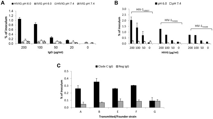
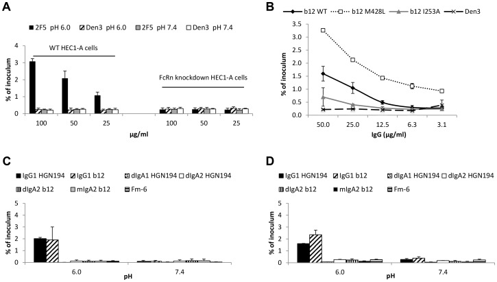
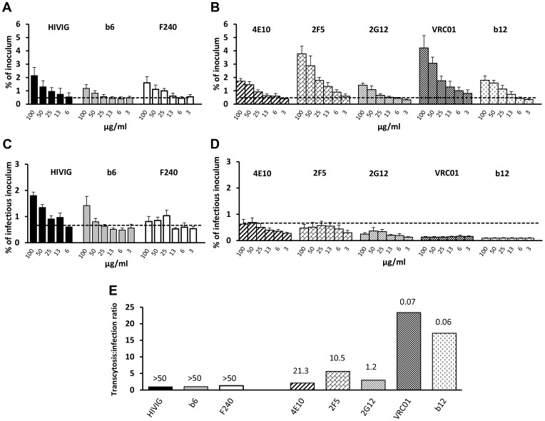
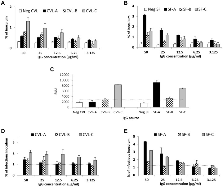
The mechanisms by which human immunodeficiency virus type 1 (HIV-1) crosses mucosal surfaces to establish infection are unknown. Acidic genital secretions of HIV-1-infected women contain HIV-1 likely coated by antibody. We found that the combination of acidic pH and Env-specific IgG, including that from cervicovaginal and seminal fluids of HIV-1-infected individuals, augmented transcytosis across epithelial cells as much as 20-fold compared with Env-specific IgG at neutral pH or non-specific IgG at either pH. Enhanced transcytosis was observed with clinical HIV-1 isolates, including transmitted/founder strains, and was eliminated in Fc neonatal receptor (FcRn)-knockdown epithelial cells. Non-neutralizing antibodies allowed similar or less transcytosis than neutralizing antibodies. However, the ratio of total:infectious virus was higher for neutralizing antibodies, indicating that they allowed transcytosis while blocking infectivity of transcytosed virus. Immunocytochemistry revealed abundant FcRn expression in columnar epithelia lining the human endocervix and penile urethra. Acidity and Env-specific IgG enhance transcytosis of virus across epithelial cells via FcRn and could facilitate translocation of virus to susceptible target cells following sexual exposure.


Conflict of interest statement

Davide Corti serves as Senior Vice President and Antonio Lanzavecchia is a scientific advisor for HUMABS BioMed SA. This does not alter our adherence to all PLOS Pathogens policies on sharing data and materials.

Figures

Figure 1
Figure 1. HIV-specific IgG and low pH enhance HIV-1 transcytosis.
(A) Indicated concentrations of HIV-specific IgG (HIVIG) or control IgG (IVIG) and 2 ng of p24 (average of 3,814,910 RNA copies) of HIV-1US712 were added to the apical surface of HEC-1A cells at pH 6.0 or 7.4. Subnatant fluid virus was quantified by RT-PCR and expressed as a percentage of inoculum applied to the apical surface of the HEC-1A cells. Data represent mean+SE of eight independent experiments (some antibody concentrations were tested less frequently); p = 3.6×10⁻⁸ comparing HIVIG at pH 6.0 with IVIG at pH 6.0; p = 3.4×10⁻⁶ comparing HIVIG at pH 6.0 with HIVIG at pH 7.4 (Kruskal-Wallis test). (B) Enhanced transcytosis occurs with multiple HIV-1 strains, including clade B R5 (HIV-1US657), R5/X4 (HIV-1HT593), and X4 (HIV-1HT599) strains. Data represent mean+SE of three independent experiments for HIV-1US657 (average inoculum = 1,096,491 RNA copies) and one experiment each for HIV-1HT593 (inoculum = 2,450,255 RNA copies) and HIV-1HT599 (inoculum = 1,477,802 RNA copies). (C) IgG enhances transcytosis of clade C transmitted/founder Env-pseudotyped virus. IgG from serum of clade C-infected or uninfected subjects was used at 50 µg/ml. Results are mean+SE of two independent experiments (average inoculum = 1,556,522 RNA copies) at pH 6.0 only; p = 0.008 comparing clade C and HIV-Neg IgG (Kruskal-Wallis test).
Figure 2
Figure 2. FcRn is responsible for pH/antibody-mediated enhanced transcytosis of HIV-1.
(A) Knockdown of FcRn eliminates enhanced transcytosis. Transcytosis was performed with the wild-type and FcRn knockdown HEC-1A cells in parallel at pH 6.0 or 7.4 using HIV-1US657 (average inoculum = 712,745 RNA copies) and mAb 2F5 or control mAb (Den3). Results represent mean+SE of three independent experiments; p = 0.032 comparing wild type and knockdown cells using 2F5 at pH 6.0 (repeated-measures ANCOVA). (B) An Fc mutant of mAb b12 that increases FcRn binding (M428L) increases transcytosis of HIV-1JRFL at pH 6.0, whereas a mutant that decreases FcRn binding (I253A) reduces transcytosis. Data represent the mean+SE of two independent experiments. (C)(D) IgG1, but not IgA1 or IgA2, enhances transcytosis at pH 6.0. mAb IgG1 b12 or its dimeric (dIgA2 b12) and monomeric (mIgA2 b12) class-switched versions and mAb IgG1 HGN194 or class switched variants dIgA1 HGN194 and dIgA2 HGN194 were tested for transcytosis using (B) HIV-1JRFL Env-pseudotyped virus (average inoculum = 2,269,529 RNA copies) or (C) SHIV1157ipEL-p (average inoculum = 682,724 RNA copies). Fm-6 is an anti-SARS coronavirus IgG1 used as a negative control. All mAbs were tested at 50 µg/ml. Data represent mean+SE of two independent assays for each virus. With either virus, IgG1 versions of the mAbs allow significantly more transcytosis at pH 6.0 than do IgA versions combined (p = 0.025 for HIV-1JRFL pseudoviruses and p = 0.021 for SHIV1157ipEL-p, Kruskal-Wallis test); there is no significant difference between IgG and IgA versions at pH 7.4 (p>0.05).
Figure 3
Figure 3. Effect of antibodies on transcytosis and infectivity.
Transcytosis (pH 6.0) of HIV-1JRFL by (A) poorly neutralizing HIVIG and mAbs b6 and F240 and (B) neutralizing mAbs 4E10, 2F5, 2G12, VRC01 and b12. HIV-1JRFL average inoculum = 3,990,745 RNA copies. Dotted lines represent mean+2 SD of % virus transcytosed with Den3 control mAb. There are significant differences between the Den3 control and 2F5 (p = 0.0002), b12 (p = 0.007), 2G12 (p = 0.006), 4E10 (p = 0.001), b6 (p = 0.008), F240 (p = 0.002), and VRC01 (p = 0.003) and between IVIG (not shown) and HIVIG (p = 0.033) (repeated-measures ANCOVA). (C) Infectivity of HIV-1JRFL whose transcytosis was mediated by poorly neutralizing or (D) neutralizing antibodies. All antibodies resulted in transcytosed virus that was infectious (>mean+2 SDs of relative light units [RLUs] from uninfected TZM-bl cells [dotted line]); average infectious inoculum = 244,601 RLUs. Compared to Den3 control, less infectious virus was transcytosed with b12 (p = 3.4×10⁻¹¹), VRC01 (p = 6.9×10⁻¹⁰), and 2G12 (p = 3.6×10⁻⁴) (repeated-measures ANCOVA). Compared to Den3, there was more infectious virus with F240 (p = 0.036) and with b6 (p = 0.068). Compared to IVIG (not shown), HIVIG resulted in more infectious virus (p = 0.003). Data in AD represent mean+SE of three or four independent experiments. (E) Ratio of % transcytosed:% infectious virus from data in (A) and (B) using 50 µg/ml of antibody. Numbers over bars represent IC50 against HIV-1JRFL.
Figure 4
Figure 4. IgG purified from genital tract secretions enhances transcytosis at pH 6.0.
(A) Cervicovaginal (CVL) fluid IgG from three HIV-infected women (CVL-A, CVL-B, and CVL-C) mediates enhanced transcytosis of HIV-1US657 (average inoculum = 2,479,412 RNA copies) at pH 6.0. (B) Similarly, IgG purified from seminal fluid of three HIV-infected men (SF-A, SF-B, and SF-C) enhances transcytosis of HIV-1US657 (average inoculum = 622,642 RNA copies) at pH 6.0. (C) Infectious HIV-1 capture by antibody in CVL and seminal fluids. Virus was captured by the indicated IgG (5 µg/ml) and added to TZM-bl cells; infection was then quantified by RLUs. Infectious virus capture correlates with transcytosis at pH 6.0 (Spearman's rho = 0.94; p = 0.005). Shown is the mean+SE of duplicate samples. The dotted line represents a negative cutoff based on the mean+2 SD of IgG from cervicovaginal or seminal fluid of HIV-uninfected subjects. (D) (E) Both cervicovaginal fluid IgG (D) and seminal fluid IgG (E) mediate transcytosis of infectious virus. Average infectious inoculum = 1,160,894 and 1,913,288 relative light units [RLUs] for cervicovaginal and seminal fluid, respectively. The dotted lines represent negative cutoffs based on the mean+2 SD of IgG from cervicovaginal and seminal fluid of the HIV-uninfected subjects. Data shown (A, B, D, and E) are mean+SE of two independent assays.
Figure 5
Figure 5. FcRn expression in human genital tissues, detected by immunohistochemistry.
FcRn expression (red stain) in columnar epithelia lining (A) the penile urethra (representative of 16 donors), (B) endocervix (representative of five donors), or (C) vagina (representative of 12 donors). Tissues were also stained with negative control (non-specific) IgG and processed through the same immunohistochemistry procedure: (D) penile urethra, (E) endocervix, and (F) vaginal epithelia. Epithelia (EP) and lamina propria (LP) are labeled. Staining was performed on paraffin-embedded specimens.

References

    1. Cohen MS (2004) HIV and sexually transmitted diseases: lethal synergy. Top HIV Med 12: 104–107. - PubMed
    1. Galvin SR, Cohen MS (2004) The role of sexually transmitted diseases in HIV transmission. Nat Rev Microbiol 2: 33–42. - PubMed
    1. Fanibunda SE, Modi DN, Gokral JS, Bandivdekar AH (2011) HIV gp120 binds to mannose receptor on vaginal epithelial cells and induces production of matrix metalloproteinases. PLoS One 6: e28014. - PMC - PubMed
    1. Mesman AW, Geijtenbeek TB (2012) Pattern Recognition Receptors in HIV Transmission. Front Immunol 3: 59. - PMC - PubMed
    1. Ballweber L, Robinson B, Kreger A, Fialkow M, Lentz G, et al. (2011) Vaginal langerhans cells nonproductively transporting HIV-1 mediate infection of T cells. J Virol 85: 13443–13447. - PMC - PubMed

Publication types

MeSH terms