. 2013 Nov 22;8(11):e79155.
doi: 10.1371/journal.pone.0079155. eCollection 2013.

The monogenean which lost its clamps

Affiliations

The monogenean which lost its clamps

Jean-Lou Justine et al. PLoS One. .

Abstract

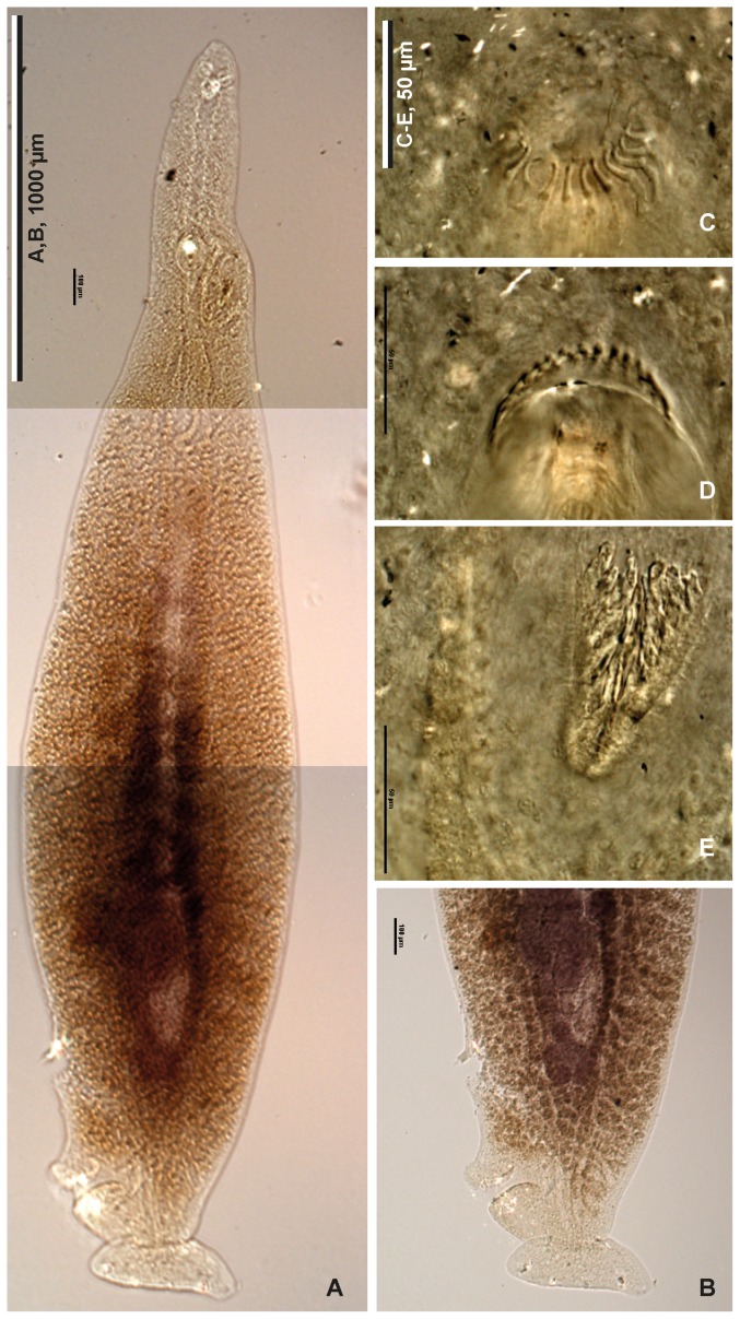
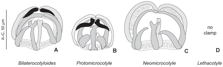
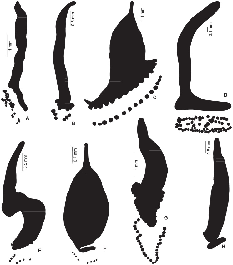
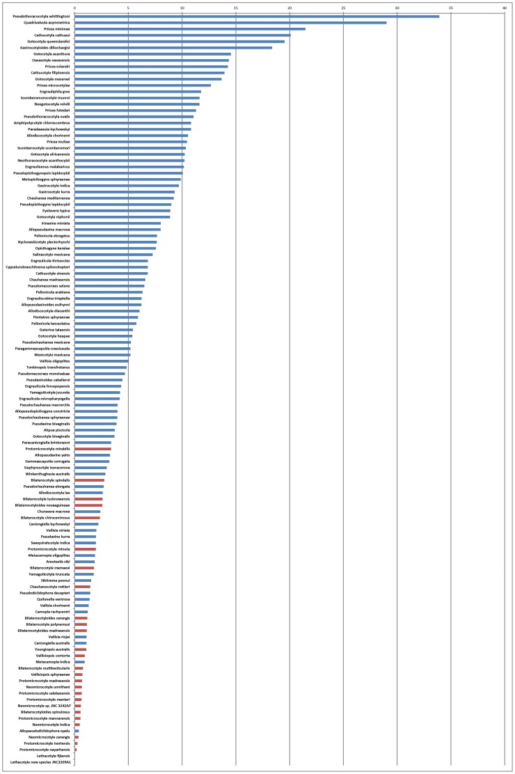
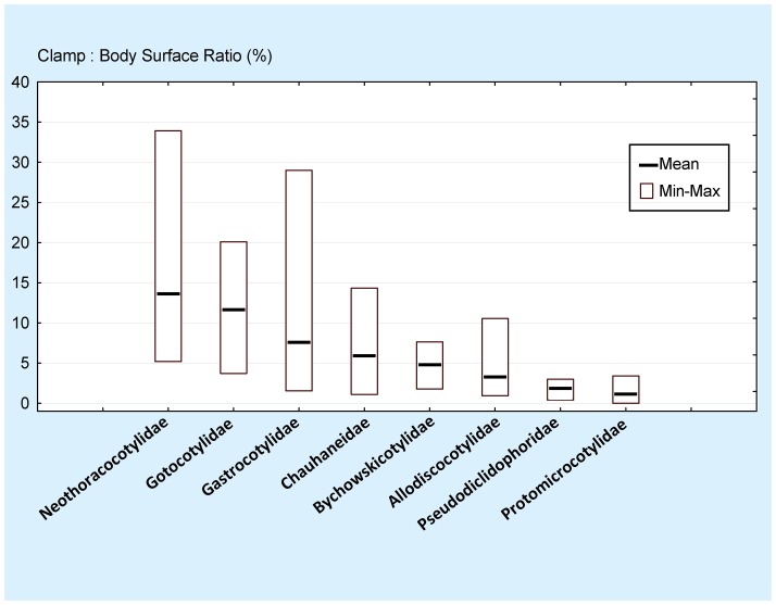
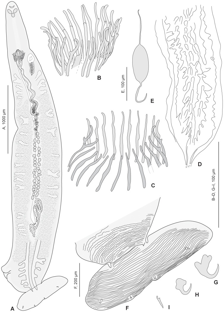
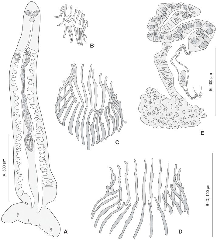
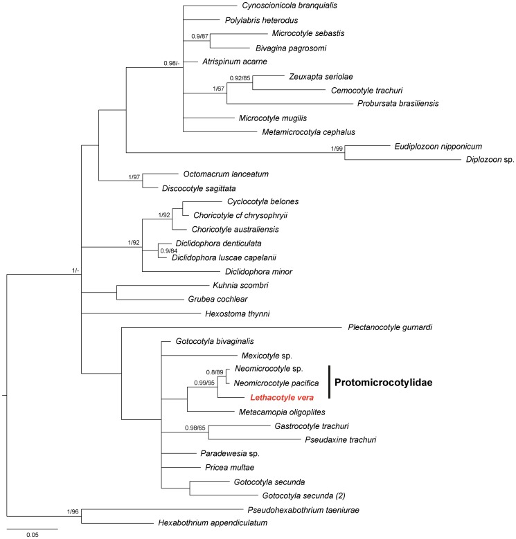
Ectoparasites face a daily challenge: to remain attached to their hosts. Polyopisthocotylean monogeneans usually attach to the surface of fish gills using highly specialized structures, the sclerotized clamps. In the original description of the protomicrocotylid species Lethacotyle fijiensis, described 60 years ago, the clamps were considered to be absent but few specimens were available and this observation was later questioned. In addition, genera within the family Protomicrocotylidae have either clamps of the "gastrocotylid" or the "microcotylid" types; this puzzled systematists because these clamp types are characteristic of distinct, major groups. Discovery of another, new, species of the genus Lethacotyle, has allowed us to explore the nature of the attachment structures in protomicrocotylids. Lethacotyle vera n. sp. is described from the gills of the carangid Caranx papuensis off New Caledonia. It is distinguished from Lethacotyle fijiensis, the only other species of the genus, by the length of the male copulatory spines. Sequences of 28S rDNA were used to build a tree, in which Lethacotyle vera grouped with other protomicrocotylids. The identity of the host fish was confirmed with COI barcodes. We observed that protomicrocotylids have specialized structures associated with their attachment organ, such as lateral flaps and transverse striations, which are not known in other monogeneans. We thus hypothesized that the clamps in protomicrocotylids were sequentially lost during evolution, coinciding with the development of other attachment structures. To test the hypothesis, we calculated the surfaces of clamps and body in 120 species of gastrocotylinean monogeneans, based on published descriptions. The ratio of clamp surface: body surface was the lowest in protomicrocotylids. We conclude that clamps in protomicrocotylids are vestigial organs, and that occurrence of "gastrocotylid" and simpler "microcotylid" clamps within the same family are steps in an evolutionary sequence, leading to the absence of these attributes in species of Lethacotyle.


Conflict of interest statement

Competing Interests: The authors have declared that no competing interests exist.

Figures

Figure 1
Figure 1. The single specimen of Lethacotyle available for study before this paper.
The slide containing the single specimen of Lethacotyle available for study before this paper: holotype of Lethacotyle fijiensis Manter & Prince, 1953 (urn:lsid:zoobank.org:act:DA367684-AAC2-44D0-A8E8-64894AFA647A), slide USNPC 48718. Our study is another example of the importance of Museum collections for modern research , .
Figure 2
Figure 2. Photograph of the holotype of Lethacotyle fijiensis Manter & Prince, 1953.
Lethacotyle fijiensis Manter & Prince, 1953 (urn:lsid:zoobank.org:act:DA367684-AAC2-44D0-A8E8-64894AFA647A). Holotype, slide USNPC 48718. A, body. B, posterior part of body, different focus. C, D, spines of male copulatory organ, two different focuses. E, sclerotised vagina. Original photographs taken by Patricia Pilitt, USNPC.
Figure 3
Figure 3. Clamps in various genera of Protomicrocotylidae.
Examples of clamps in various genera of Protomicrocotylidae. A, Bilaterocotyloides novaeguineae (Rohde, 1977) Lebedev, 1986 (USNPC 74800). B, Protomicrocotyle sp. (MNHN JNC1163A5). C, Neomicrocotyle sp. (MNHN JNC3242A4). Black: additional sclerite, characteristic of the “gastrocotylid” clamp. Bilaterocotyloides and Protomicrocotyle have clamps of the “gastrocotylid” type, Neomicrocotyle has clamps of the “microcotylid” type, and Lethacotyle has no clamp.
Figure 4
Figure 4. Body and clamp surfaces: examples of line drawings in 8 families.
Body and clamp surfaces: examples of line drawings used for data extraction in each of the 8 families of the Gastrocotylinea. All species drawn to same body length. A, Gotocotylidae, Gotocotyla niphonii. B, Bychowskicotylidae, Tonkinopsis transfretanus. C, Gastrocotylidae, Allopseudaxinoides euthynni. D, Neothoracocotylidae, Pricea minimae. E, Allodiscocotylidae, Metacamopia indica. F, Pseudodiclidophoridae, Allopseudodiclidophora opelu. G, Chauhaneidae, Cotyloatlantica mediterranea. H, Protomicrocotylidae, Lethacotyle vera n. sp (no clamps). Details in Table 3.
Figure 5
Figure 5. Ratio between clamp surface and body surface in species of gastrocotylinean monogeneans.
Ratio between clamp surface and body surface in species of gastrocotylinean monogeneans. Ratios are ordered in decreasing sequence. Red: protomicrocotylids; blue: species of other families.
Figure 6
Figure 6. Ratio between clamp surface and body surface in families of gastrocotylinean monogeneans.
Ratio between clamp surface and body surface in families of gastrocotylinean monogeneans. Ratios are ordered in decreasing order of mean. Protomicrocotylids have the lowest mean and lowest minimum. For significance see Table 4.
Figure 7
Figure 7. Lethacotyle vera n. sp. Adult and details.
Lethacotyle vera n. sp (urn:lsid:zoobank.org:act:0B7ABE99-07AF-4088-97F3-1A154DBA614D). A, whole body; B, spines of male copulatory organ (MCO). C, spines of MCO in other specimen (paratype MNHN JNC1189A3). D, sclerotized vagina. E, egg, in utero. F, striations on posterior part of body; G, H, I, hooks (paratype MNHN JNC1185A3). A, B, D, F: holotype, MNHN JNC3209A1.
Figure 8
Figure 8. Lethacotyle vera n. sp. Juvenile and other details.
Lethacotyle vera n. sp (urn:lsid:zoobank.org:act:0B7ABE99-07AF-4088-97F3-1A154DBA614D). A, juvenile (specimen MNHN JNC3188A1). B, spines of MCO in juvenile. C, spines of MCO in paratype MNHN JNC3188A2c (posterior part of body processed for molecular study); D, spines of MCO in paratype MNHN JNC1189A2. E, outline of ovary (paratype JNC1189A1).
Figure 9
Figure 9. Tree of gastrocotylinean monogeneans.
Tree of gastrocotylinean monogeneans, based on a phylogenetic analysis of 28S sequences.

References

    1. Justine J-L (1998) Non-monophyly of the monogeneans? International Journal for Parasitology 28: 1653–1657. - PubMed
    1. Perkins EM, Donnellan SC, Bertozzi T, Whittington ID (2010) Closing the mitochondrial circle on paraphyly of the Monogenea (Platyhelminthes) infers evolution in the diet of parasitic flatworms. International Journal for Parasitology 40: 1237–1245. - PubMed
    1. Mollaret I, Jamieson BGM, Adlard RD, Hugall A, Lecointre G, et al. (1997) Phylogenetic analysis of the Monogenea and their relationships with Digenea and Eucestoda inferred from 28S rDNA sequences. Molecular and Biochemical Parasitology 90: 433–438. - PubMed
    1. Mollaret I, Jamieson BGM, Justine J-L (2000) Phylogeny of the Monopisthocotylea and Polyopisthocotylea (Platyhelminthes) inferred from 28S rDNA sequences. International Journal for Parasitology 30: 171–185. - PubMed
    1. Littlewood DTJ, Rohde K, Clough KA (1999) The interrelationships of all major groups of Platyhelminthes: phylogenetic evidence from morphology and molecules. Biological Journal of the Linnean Society 66: 75–114.

Publication types

LinkOut - more resources