. 2013 Nov 22;8(11):e79919.
doi: 10.1371/journal.pone.0079919. eCollection 2013.

IQ domain GTPase-activating protein 1 is involved in shear stress-induced progenitor-derived endothelial cell alignment

Affiliations

IQ domain GTPase-activating protein 1 is involved in shear stress-induced progenitor-derived endothelial cell alignment

Lila Rami et al. PLoS One. .

Abstract

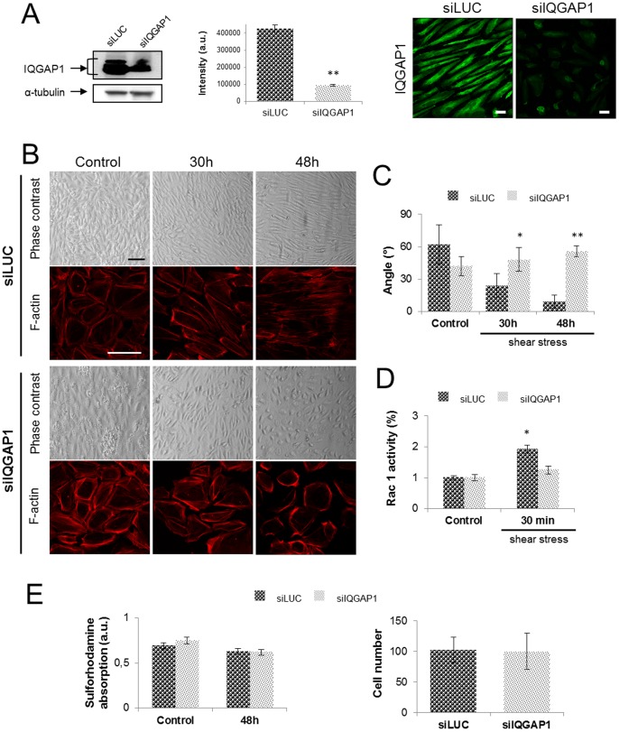
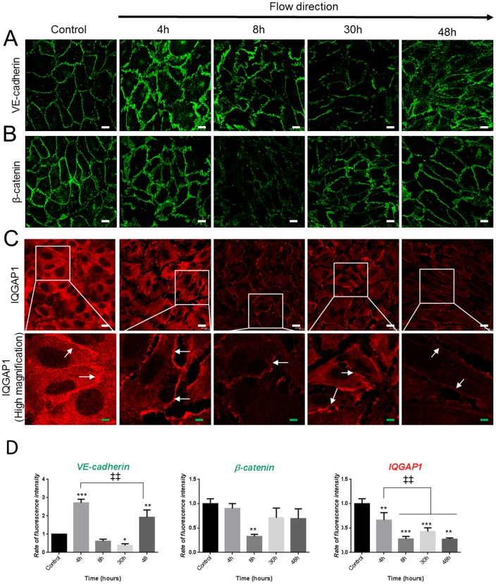
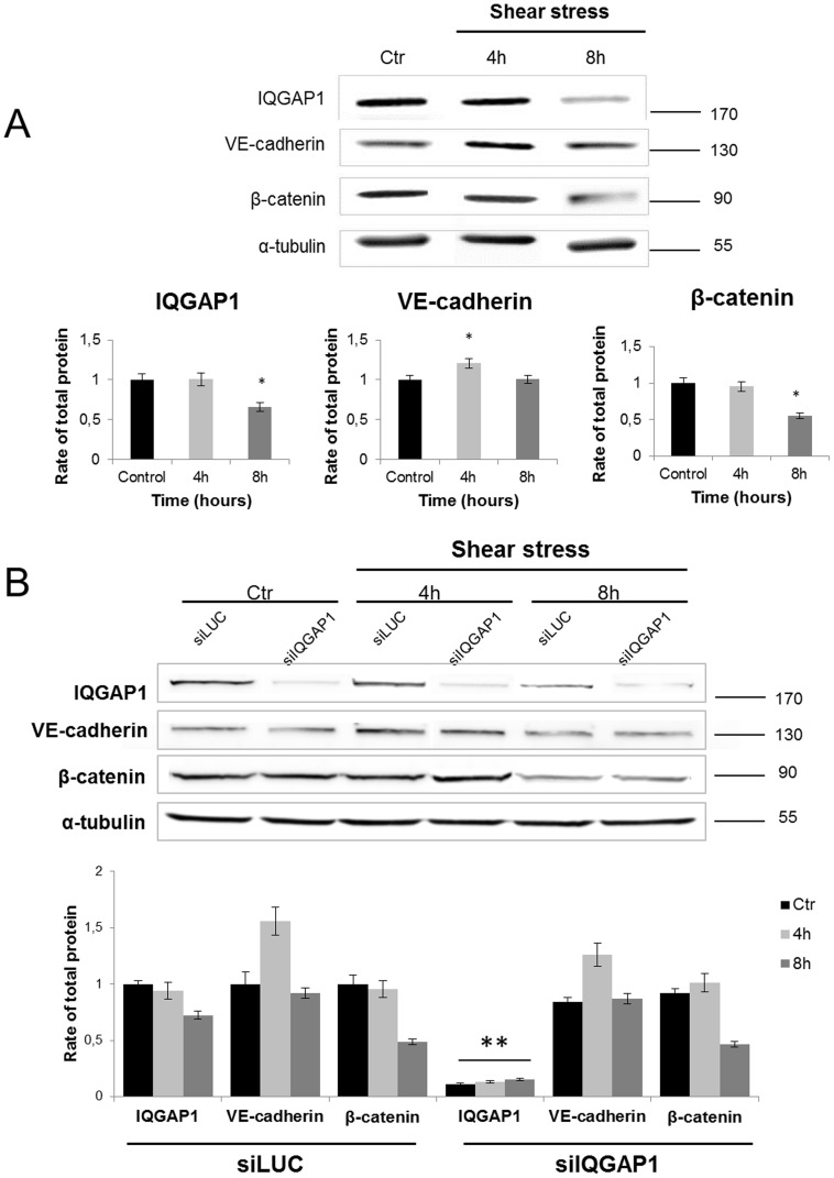
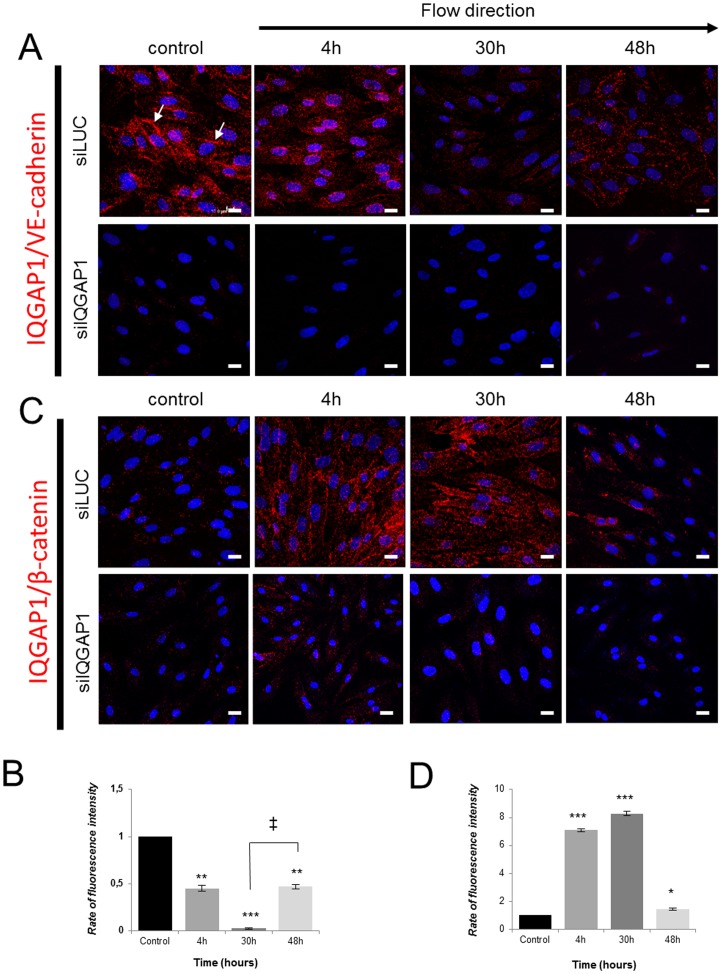
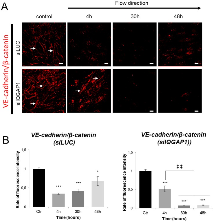
Shear stress is one of mechanical constraints which are exerted by blood flow on endothelial cells (ECs). To adapt to shear stress, ECs align in the direction of flow through adherens junction (AJ) remodeling. However, mechanisms regulating ECs alignment under shear stress are poorly understood. The scaffold protein IQ domain GTPase activating protein 1 (IQGAP1) is a scaffold protein which couples cell signaling to the actin and microtubule cytoskeletons and is involved in cell migration and adhesion. IQGAP1 also plays a role in AJ organization in epithelial cells. In this study, we investigated the potential IQGAP1 involvement in the endothelial cells alignment under shear stress. Progenitor-derived endothelial cells (PDECs), transfected (or not) with IQGAP1 small interfering RNA, were exposed to a laminar shear stress (1.2 N/m(2)) and AJ proteins (VE-cadherin and β-catenin) and IQGAP1 were labeled by immunofluorescence. We show that IQGAP1 is essential for ECs alignment under shear stress. We studied the role of IQGAP1 in AJs remodeling of PDECs exposed to shear stress by studying cell localization and IQGAP1 interactions with VE-cadherin and β-catenin by immunofluorescence and Proximity Ligation Assays. In static conditions, IQGAP1 interacts with VE-cadherin but not with β-catenin at the cell membrane. Under shear stress, IQGAP1 lost its interaction from VE-cadherin to β-catenin. This "switch" was concomitant with the loss of β-catenin/VE-cadherin interaction at the cell membrane. This work shows that IQGAP1 is essential to ECs alignment under shear stress and that AJ remodeling represents one of the mechanisms involved. These results provide a new approach to understand ECs alignment under to shear stress.


Conflict of interest statement

Competing Interests: The authors have declared that no competing interests exist.

Figures

Figure 1
Figure 1. PDECs align in the direction of flow under shear stress.
(A) PDECs exposed to static or shear stress conditions (1.2 N/m2) were observed after fixation by optical microscopy. Cells not exposed to flow were used as control. Progressively, PDECs organized in a monolayer of contiguous cells, detached each other (arrows at 4 h) and aligned in the direction of flow (Scale bar: 100 µm). In lower panel, the actin cytoskeleton was labeled in PDECs exposed or not to shear stress, showing cortical actin and stress fiber formation within cells. (B) Cells direction has been quantified under static and shear stress conditions using the ImageJ software. The vertical axis and circular arcs corresponding to the standard error of mean (S.E.M.) and the horizontal axis representing the flow direction.
Figure 2
Figure 2. IQGAP1 is necessary for cell-cell adhesion and PDECs alignment.
The IQGAP1 extinction rate has been evaluated by immunofluorescence staining of IQGAP1 and western blot before (not shown) and after 48 h of exposition to shear stress (scale bar: 10 µm) (A). PDECs were transfected with siRNA against IQGAP1 (siIQGAP1) or Luciferase (siLUC, control), exposed to shear stress for the indicated times (scale bar: 100 µm) and F-actin was stained with coupled-TRITC phalloïdine antibody (scale bar: 100 µm) (B). Angles of siLUC or siIQGAP1 transfected PDECs orientation under shear stress have been measured for 3 independent experiments and are represented in (C). The Rac1 activity has been quantified for PDECs transfected with siLUC or siIQGAP1 and maintained in static conditions or exposed to shear stress for 30 h and 48 h, showing the non Rac1 activation for siIQGAP1-transfected PDECs (D). Cell viability has been checked by Sulforhodamine test and cell number has been quantified in static conditions (control) and after 48 h of shear stress (E).
Figure 3
Figure 3. Shear stress induces the adherens junction remodeling (A and B) and IQGAP1 delocalization (C).
VE-cadherin, β-catenin and IQGAP1 immunofluorescence staining in control conditions and after flow are represented in A, B and C respectively. Fluorescence intensities at the cell membrane are quantified in (D) and total protein expression were assessed by western blot for cells maintained in static conditions (control) or exposed to shear stress for 8 h (E). (*p<0.05; **p<0.01; ***p<0.001 refer to comparison with control conditions. ‡‡p<0.01 refer to indicated comparisons) (white scale bar: 10 µm; green scale bar: 3 µm).
Figure 4
Figure 4. Total protein expression of IQGAP1, VE-cadherin and b-catenin in PDECs transfected or not with siRNA, before and after shear stress exposure.
The total expression of IQGA1, VE-cadherin and β-catenin in non-transfected PDECs was investigated by western blot in static conditions (Ctr) and after 4 h and 8 h of shear stress, and quantified in comparison with the static condition (Ctr) (*p<0.05) (A). The same experiment was performed on siRNA (LUC or IQGAP1) transfected cells (**p<0.01 refer to comparison with siLUC conditions) (B).
Figure 5
Figure 5. IQGAP1 downregulation induces the lack of cell cohesiveness.
In (A) are represented immunofluorescence staining of VE-cadherin and β-catenin in PDECs transfected with siIQGAP1 or siLUC, under static conditions. VE-cadherin and β-catenin expression were discontinuous at the cell membrane in siIQGAP1-transfected PDECs (arrows). Arrowheads show lacunas formed by the adherens junction weakness (Scale bar = 100 µm). Permeability assay was performed by quantification of FITC-dextran (70 kDa) passage through a monolayer of not transfected or siRNA (LUC or IQGAP1) -transfected PDECs seeded in a Transwell®. The transfection with siIQGAP1 seems to increase the monolayer permeability (B). PDECs: untransfected cells; siIQGAP1: IQGAP1 siRNA-transfected cells; siLUC: Luciferase siRNA-transfected cells; Dextran free: serum free medium without dextran (negative control); Dextran only: dextran-FITC added to serum free medium without cells.
Figure 6
Figure 6. IQGAP1 and adherens junction proteins interactions are modulated under shear conditions.
PDECs are exposed to shear stress and interactions between IQGAP1 and VE-cadherin (A) and IQGAP1 and β-catenin (B) were analyzed with the Duolink™ kit. Proteins interactions at cell membrane are showed by arrows. (B) and (D) show the rate of fluorescence intensity (normalized to that of control) at the cell membrane, after shear stress for IQGAP1/VE-cadherin and IQGAP1/β-catenin interactions, respectively. (*p<0.05; **p<0.01; ***p<0.001 refer to comparison with control conditions. p<0.05 refers to indicated comparisons) (Scale bar: 10 µm).
Figure 7
Figure 7. VE-cadherin and β-catenin interactions are modulated under shear conditions.
PDECs are exposed to shear stress and interactions between VE-cadherin and β-catenin, were labeled with the Duolink kit in cells transfected with either siLUC or siIQGAP1 (A). Proteins interactions at cell membrane are showed by arrows. B and C show the rate of fluorescence intensity (normalized to that of control) after shear stress for VE-cadherin/β-catenin interaction for siLUC or siIQGAP1 transfected cells, respectively. (*p<0.05; **p<0.01; ***p<0.001 refer to comparison with control conditions. p<0.05 refers to indicated comparisons) (Scale bar: 10 µm).

References

    1. Noria S, Cowan DB, Gotlieb AI, Langille BL (1999) Transient and steady-state effects of shear stress on endothelial cell adherens junctions. Circ Res 85: 504–514. - PubMed
    1. Yamamoto K, Takahashi T, Asahara T, Ohura N, Sokabe T, et al. (2003) Proliferation, differentiation, and tube formation by endothelial progenitor cells in response to shear stress. J Appl Physiol 95: 2081–2088. - PubMed
    1. Ukropec JA, Hollinger MK, Woolkalis MJ (2002) Regulation of VE-cadherin linkage to the cytoskeleton in endothelial cells exposed to fluid shear stress. Exp Cell Res 273: 240–247. - PubMed
    1. Resnick N, Yahav H, Shay-Salit A, Shushy M, Schubert S, et al. (2003) Fluid shear stress and the vascular endothelium: for better and for worse. Prog Biophys Mol Biol 81: 177–199. - PubMed
    1. Tzima E, Irani-Tehrani M, Kiosses WB, Dejana E, Schultz DA, et al. (2005) A mechanosensory complex that mediates the endothelial cell response to fluid shear stress. Nature 437: 426–431. - PubMed

Publication types

MeSH terms