. 2013 Nov 21;8(11):e80153.
doi: 10.1371/journal.pone.0080153. eCollection 2013.

Heparin modulates the endopeptidase activity of Leishmania mexicana cysteine protease cathepsin L-Like rCPB2.8

Affiliations

Heparin modulates the endopeptidase activity of Leishmania mexicana cysteine protease cathepsin L-Like rCPB2.8

Wagner A S Judice et al. PLoS One. .

Abstract

Background: Cysteine protease B is considered crucial for the survival and infectivity of the Leishmania in its human host. Several microorganism pathogens bind to the heparin-like glycosaminoglycans chains of proteoglycans at host-cell surface to promote their attachment and internalization. Here, we have investigated the influence of heparin upon Leishmania mexicana cysteine protease rCPB2.8 activity.

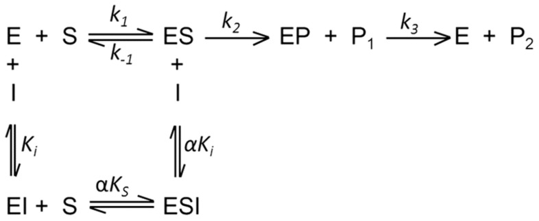
Methodology/principal findings: THE DATA ANALYSIS REVEALED THAT THE PRESENCE OF HEPARIN AFFECTS ALL STEPS OF THE ENZYME REACTION: (i) it decreases 3.5-fold the k 1 and 4.0-fold the k -1, (ii) it affects the acyl-enzyme accumulation with pronounced decrease in k 2 (2.7-fold), and also decrease in k 3 (3.5-fold). The large values of ΔG = 12 kJ/mol for the association and dissociation steps indicate substantial structural strains linked to the formation/dissociation of the ES complex in the presence of heparin, which underscore a conformational change that prevents the diffusion of substrate in the rCPB2.8 active site. Binding to heparin also significantly decreases the α-helix content of the rCPB2.8 and perturbs the intrinsic fluorescence emission of the enzyme. The data strongly suggest that heparin is altering the ionization of catalytic (Cys(25))-S(-)/(His(163))-Im(+) H ion pair of the rCPB2.8. Moreover, the interaction of heparin with the N-terminal pro-region of rCPB2.8 significantly decreased its inhibitory activity against the mature enzyme.

Conclusions/significance: Taken together, depending on their concentration, heparin-like glycosaminoglycans can either stimulate or antagonize the activity of cysteine protease B enzymes during parasite infection, suggesting that this glycoconjugate can anchor parasite cysteine protease at host cell surface.


Conflict of interest statement

Competing Interests: The authors have declared that no competing interests exist.

Figures

Figure 1
Figure 1. Schematic mechanism of hydrolytic process of a cysteine protease and inhibition.
Individual constants k 1, k −1, k 2 and k 3 of the hydrolytic reactions of cysteine protease. k1 is the substrate diffusion constant into the active site, k–1 the substrate dissociation constant, k2 the acylation constant, k3 the deacylation constant, KS  =  (k 1 +k2)/k1, K i the inhibition constant, and α is the parameter of K S and K i perturbation.
Figure 2
Figure 2. Effect of Heparin upon the rCPB2.8 Endopeptidase Activity.
The influence of heparin upon rCPB2.8 endopeptidase activity was monitored fluorometrically using the fluorogenic substrate Z-FR-MCA. The steady-state kinetic assays with fluorogenic substrate were performed in 100 mM sodium acetate buffer (pH 5.5) containing 20% glycerol and 5 mM DTT at 35°C. The enzyme was activated by its pre-incubation in the assay buffer for 5 min at 35°C before the substrate addition. A, the rate of substrate Z-FR-MCA hydrolysis as a function of substrate concentration. The kinetic parameters were determined by measuring the initial rate of hydrolysis at various substrate concentrations in the absence or in the presence of heparin, control (▵–▵); 8 µM (•–•); 16 µM (•–•); 32 µM (□–□); 42 µM (▴–▴); 48 µM (○–○); 62 µM (*–*) of heparin. B , the reciprocal plot 1/V versus 1/[S] in the presence of different concentrations of heparin (0 — 62 µM). C, replots of slope 1/[S] versus [heparin]. D, replot of 1/V −axis intercept versus [heparin]. The data of replots were taken from the reciprocal plot 1/V versus 1/[S].
Figure 3
Figure 3. Arrhenius and Eyring plots of the kcat/KM and kcat for the Z-FR-MCA hydrolisys by rCPB2.8.
A, Arrhenius plot of the ln (kcat/K M ) versus 1000/T. B, Arrhenius plot of the ln k cat versus 1000/T. C, Eyring plot of the ln (kcat/K M.T) versus 1000/T. D, Eyring plot of the ln (kcat/T) versus 1000/T. The experimental conditions were: the values of the kinetic parameters kcat and K M for rCPB2.8 was determined using 100 mM sodium acetate buffer, 20% glycerol, 5 mM DTT, pH 5.5 activating the enzyme for 10 min in the temperature range 10°C to 55°C as described under “Methods.” The data were obtained in the absence (•–•) or in the presence (○–○) of 40 µM heparin. The continuous lines (slops) were drawn according the Equations 6, 7, 9 and 10, with the best-fit parameter values listed in Table 1 and 2.
Figure 4
Figure 4. The influence of heparin upon rCPB2.8 pH activity profiles
. The substrate Z-FR-MCA hydrolysis was performed in absence (•–•) and in presence (○–○) of 40 µM heparin at 35°C in universal buffer containing 25 mM glycine, 25 mM acetic acid, 25 mM Mes and 75 mM Tris, containing 20% glycerol and 5 mM DTT, the pH of buffers were adjusted using HCl or NaOH diluted solutions as described under “Methods.” A, the pH activity profiles data were fitted according to Equation 10 by using non-linear regression software system. The associated table shows the values of pK ES1 and pK ES2 in absence and in the presence of heparin. B, Dixon-Web plot of the log V maxapp versus pH where the dot lines of slope  =  1 and slope  =  -1 tangent to the curve.
Figure 5
Figure 5. Heparin binding-induced rCPB2.8 conformational change.
A, the intrinsic fluorescence of tryptophan residues of the rCPB2.8 was monitored in 50 mM sodium phosphate buffer containing 20% glycerol at 35 °C by measuring the emission of fluorescence between at 300 — 450 nm after excitation at λex  =  290 nm in the absence or in the presence of different concentrations of heparin (0 — 167 µM). The insert in A shows the variation of the intrinsic fluoresce emission ΔF (F-F0) as a function of heparin concentration. B, effects of heparin on rCPB2.8 circular dichroism spectra. About 2 µM rCPB2.8 were determined in 5 mM sodium phosphate buffer pH 5.8 in the absence (•–•) or in the presence (□–□) of 40 µM heparin.
Figure 6
Figure 6. Effects of heparin on the N-terminal pro-region of rCPB2.8.
A, the influence of heparin concentration upon N-terminal pro-region segment intrinsic fluorescence emission. The insert in A shows the variation of the intrinsic fluoresce emission ΔF (F-F0) of the N-terminal pro-region as a function of heparin concentration (0 — 167 µM) as described under “Methods.” B, heparin prevents the inhibitory activity of N-terminal pro-region upon rCPB2.8. The remaining activity of the enzyme was plotted as a function of N-terminal pro-region concentration in the absence (○–○) or in the presence (•–•) of 40 µM heparin.

References

    1. Sajid M, McKerrow JH (2002) Cysteine proteases of parasitic organisms. Mol Biochem Parasitol 120: 1-21. Review. Erratum in: Mol Biochem Parasitol 121: 159. - PubMed
    1. Coombs GH (1982) Proteinases of Leishmania mexicana and other flagellate protozoa. Parasitology 84: 149–155. - PubMed
    1. Pupkis MF, Coombs GH (1984) Purification and characterization of proteolytic enzymes of Leishmania mexicana amastigotes and promastigotes. J Gen Microbiol 130: 2375–2383. - PubMed
    1. Robertson CD, Coombs GH (1990) Characterization of three groups of cysteine proteinases in the amastigotes of Leishmania mexicana mexicana . Mol Biochem Parasitol 42: 269–276. - PubMed
    1. Robertson CD, Coombs GH (1992) Stage-specific proteinases of Leishmania mexicana promastigotes. FEMS Microbiol Lett 94: 127–132. - PubMed

Publication types