

Imaging findings of primary splenic lymphoma: a review of 17 cases in which diagnosis was made at splenectomy
- PMID: 24278265
- PMCID: PMC3837000
- DOI: 10.1371/journal.pone.0080264
Imaging findings of primary splenic lymphoma: a review of 17 cases in which diagnosis was made at splenectomy
Abstract
Purpose: This study sought to characterize the imaging features of primary splenic lymphoma (PSL).
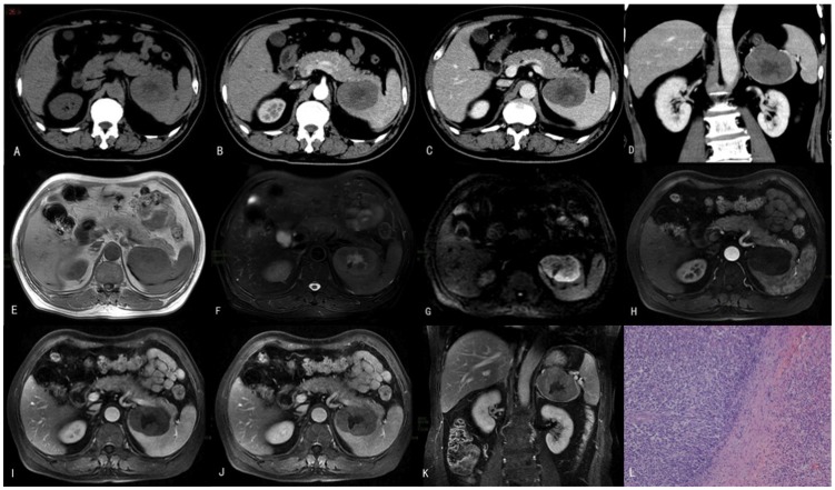
Materials and methods: Pathological and imaging data from 17 patients with primary splenic lymphoma initially diagnosed at splenectomy were retrospectively analyzed. Pretreatment computed tomography (CT) imaging was available for 16 patients, and magnetic resonance imaging (MRI) data were available for 4 patients. Splenic lymphoma imaging data were categorized based on the gross pathological presentation in the following manner: type 1, homogeneous enlargement; type 2, miliary nodules; type 3, multifocal masses of varying size; and type 4, solitary large mass.
Results: Of the 17 patients with PSL, 16 cases were non-Hodgkin lymphoma, and of these, 9 cases were diffuse large B cell lymphomas (DLBCL) and 4 cases were splenic marginal zone B-cell lymphoma (SMZL). Imaging showed the following types of PSL presentation: 1 case of type 1, 0 cases of type 2, 4 cases of type 3, and 12 cases of type 4. There was evidence of necrosis in 12 cases (70.6%), and there was evidence of mild enhancement in enhanced CT in 14 cases and in enhanced MRI in 3 cases. Prior to surgery, PSL was considered possible in 8 patients.
Conclusion: The most frequent histological subtype was DLBCL, followed by SMZL. In both CT and MRI, PSL generally presents as a solitary mass or masses rather than as splenomegaly. In addition, necrosis and mild enhancement are commonly observed, and splenectomy may be required to confirm the diagnosis.
Conflict of interest statement
Figures
Similar articles
-
Primary non-Hodgkin's splenic lymphoma.Clin Radiol. 1998 Feb;53(2):137-42. doi: 10.1016/s0009-9260(98)80061-5. Clin Radiol. 1998. PMID: 9502091
-
[Tumors of lymphoid and hematopoietic tissue of spleen: a clinicopathologic analysis of 53 cases].Zhonghua Bing Li Xue Za Zhi. 2017 Nov 8;46(11):775-781. doi: 10.3760/cma.j.issn.0529-5807.2017.11.008. Zhonghua Bing Li Xue Za Zhi. 2017. PMID: 29136691 Chinese.
-
[Splenic marginal zone lymphoma].Pan Afr Med J. 2017 Mar 1;26:111. doi: 10.11604/pamj.2017.26.111.11446. eCollection 2017. Pan Afr Med J. 2017. PMID: 28533834 Free PMC article. French.
-
Splenic marginal zone lymphoma: a case report and literature review.World J Surg Oncol. 2020 Oct 1;18(1):259. doi: 10.1186/s12957-020-02030-3. World J Surg Oncol. 2020. PMID: 33004051 Free PMC article. Review.
-
Primary splenic lymphoma.Cancer Surv. 1997;30:193-212. Cancer Surv. 1997. PMID: 9547993 Review.
Cited by
-
Multimodality imaging of spleen involvement in Erdheim-Chester disease mimicking splenic hemangioma: a unique case report.Am J Nucl Med Mol Imaging. 2023 Jun 25;13(3):118-125. eCollection 2023. Am J Nucl Med Mol Imaging. 2023. PMID: 37457326 Free PMC article.
-
Splenic Lymphomas: A Tertiary Care Center Experience and Review of Literature.Indian J Hematol Blood Transfus. 2023 Jul;39(3):402-412. doi: 10.1007/s12288-022-01621-2. Epub 2022 Dec 28. Indian J Hematol Blood Transfus. 2023. PMID: 37304493 Free PMC article.
-
Machine Learning Radiomics Signature for Differentiating Lymphoma versus Benign Splenomegaly on CT.Diagnostics (Basel). 2023 Dec 8;13(24):3632. doi: 10.3390/diagnostics13243632. Diagnostics (Basel). 2023. PMID: 38132216 Free PMC article.
-
Diagnostic performance of different imaging modalities for splenic malignancies: A comparative meta-analysis.Eur J Radiol Open. 2024 Apr 22;12:100566. doi: 10.1016/j.ejro.2024.100566. eCollection 2024 Jun. Eur J Radiol Open. 2024. PMID: 38681661 Free PMC article.
-
Optimal index for detecting splenic involvement on 18F-fluorodeoxyglucose positron emission tomography/computed tomography imaging in diffuse large B-cell lymphoma.Medicine (Baltimore). 2024 Mar 1;103(9):e37290. doi: 10.1097/MD.0000000000037290. Medicine (Baltimore). 2024. PMID: 38428864 Free PMC article.
References
-
- Ahmann DL, Kiely JM, Harrison EG Jr, Payne WS (1966) Malignant lymphoma of the spleen. A review of 49 cases in which the diagnosis was made at splenectomy. Cancer 19: 461–469. - PubMed
-
- Swerdlow SH, Campo E, Harris NL, Jaffe ES, Pileri SA, et al... (2008) World Health Organization classification of tumors of haematopoietic and lymphoid tissues. lyon, France: IARC Press.
-
- Dasgupta T, Coombes B, Brasfield RD (1965) Primary Malignant Neoplasms of the Spleen. Surg Gynecol Obstet 120: 947–960. - PubMed
Publication types
MeSH terms
LinkOut - more resources
Full Text Sources
Other Literature Sources
Medical
