. 2013 Nov 20;8(11):e80477.
doi: 10.1371/journal.pone.0080477. eCollection 2013.

Changes in the inflammatory response to injury and its resolution during the loss of regenerative capacity in developing Xenopus limbs

Affiliations

Changes in the inflammatory response to injury and its resolution during the loss of regenerative capacity in developing Xenopus limbs

Anthony L Mescher et al. PLoS One. .

Abstract

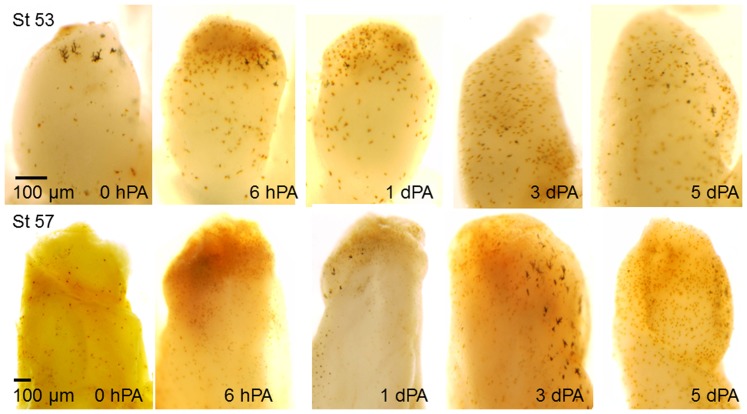
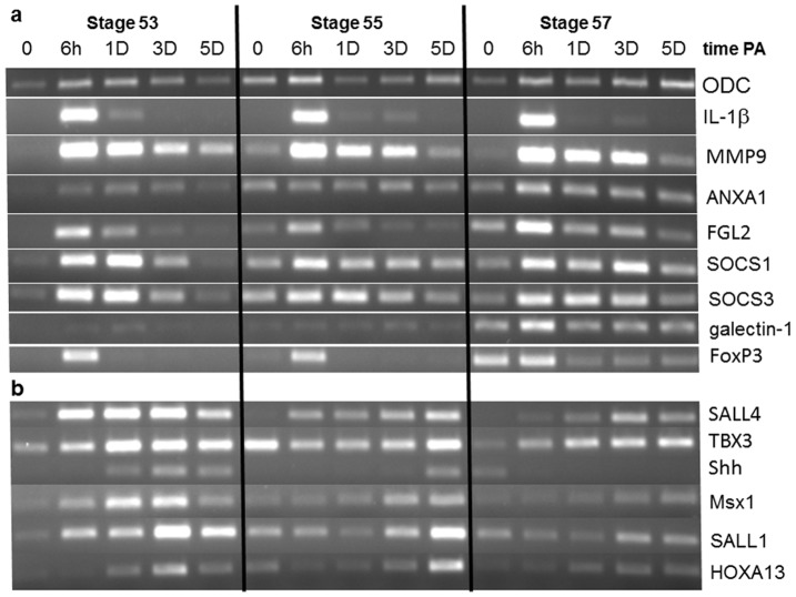
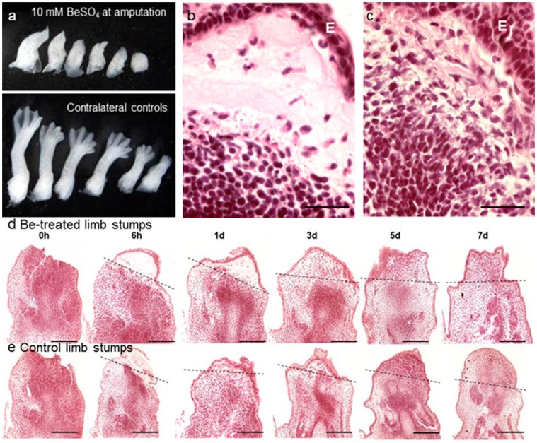
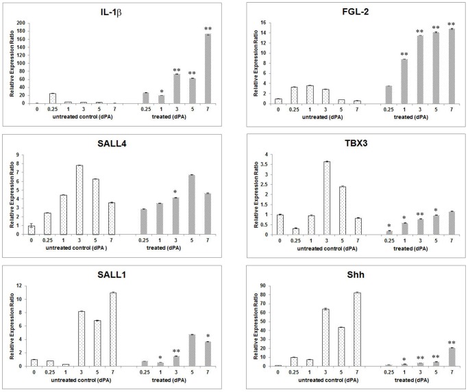
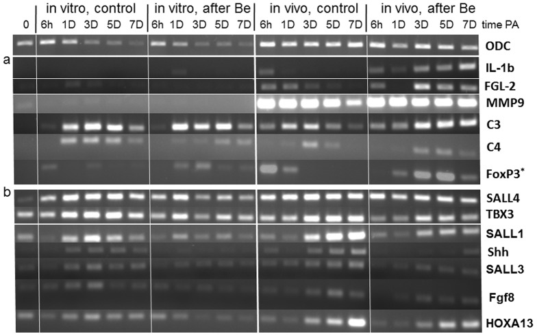
Tissue and organ regeneration, unlike development, involves an injury that in postembryonic animals triggers inflammation followed by resolution. How inflammation affects epimorphic regeneration is largely uninvestigated. Here we examine inflammation and its resolution in Xenopus laevis hindlimb regeneration, which declines during larval development. During the first 5 days postamputation, both regeneration-competent stage 53 and regeneration-deficient stage 57 hindlimbs showed very rapid accumulation of leukocytes and cells expressing interleukin-1β and matrix metalloproteinase 9. Expression of genes for factors mediating inflammatory resolution appeared more persistent at stages 55 and 57 than at stage 53, suggesting changes in this process during development. FoxP3, a marker for regulatory T cells, was upregulated by amputation in limbs at all three stages but only persisted at stage 57, when it was also detected before amputation. Expression of genes for cellular reprogramming, such as SALL4, was upregulated in limbs at all 3 stages, but markers of limb patterning, such as Shh, were expressed later and less actively after amputation in regeneration-deficient limbs. Topical application of specific proinflammatory agents to freshly amputated limbs increased interleukin-1β expression locally. With aqueous solutions of the proinflammatory metal beryllium sulfate, this effect persisted through 7 days postamputation and was accompanied by inhibition of regeneration. In BeSO4-treated limbs expression of markers for both inflammation and resolution, including FoxP3, was prolonged, while genes for cellular reprogramming were relatively unaffected and those for limb patterning failed to be expressed normally. These data imply that in Xenopus hindlimbs postamputation inflammation and its resolution change during development, with little effect on cellular dedifferentiation or reprogramming, but potentially interfering with the expression of genes required for blastema patterning. The results suggest that developmental changes in the larval anuran immune system may be involved in the ontogenetic loss of epimorphic regeneration in this system.


Conflict of interest statement

Competing Interests: The authors have declared that no competing interests exist.

Figures

Figure 1
Figure 1. Whole-mounts of stage 53 and stage 57 Xenopus hindlimbs from 0 hours to 5 days post-amputation (PA) stained by enzyme histochemistry for myeloperoxidase (MPO), a marker for neutrophils and macrophages.
Only background staining is visible at the time of amputation (0 hPA), but by 6 hPA in limbs of both stages the number of brown MPO+ cells increase greatly near the site of amputation. MPO staining diminished slowly through 5 dPA in limbs of both stages. The larger, very dark structures are melanocytes.
Figure 2
Figure 2. Expression of genes previously identified in studies of gene activity during the early phase of Xenopus limb regeneration, shown by RT-PCR at different times PA during the transition from regeneration-complete to –incomplete in stages 53, 55, and 57.
Tissue from 20 limb stumps was used for each time point and expression of the loading control ornithine decarboxylase (ODC) is shown in the top row. (a) Genes with primarily inflammation-related function: Expression of interleukin-1β (IL-1β) appeared maximal by 6 hPA but then was rapidly diminished at all stages. Expression of factors involved in the resolution of inflammation, including annexin-A1 (ANXA1), fibrinogen-like protein 2 (FGL2), suppressors of cytokine signaling (SOCS) 1 and 3, galectin-1, and the key marker for regulatory T cells, FoxP3, were also all up-regulated by 6hPA and then diminished in limbs of all 3 stages, but more slowly in regeneration-incomplete limbs suggesting that inflammation-related activity persists after amputation in the more developed limbs. Low expression of many of these pro-resolution factors, including FoxP3, was seen already at the time of amputation in regeneration-deficient limbs, indicating the presence of immune cells not found in limbs at early regeneration-complete stages. (b) Genes with primarily cell reprogramming and organ patterning function: Expression of SALL4 and TBX3, both involved in cell reprogramming during early blastema formation, are up-regulated by amputation at all 3 stages, but much more slowly and at apparently lower levels in regeneration-incomplete limbs. Expression of genes required for blastema patterning, including Shh, Msx1, SALL1, and HOXA13, occurred at various times PA in stage 53 limbs, but was increasingly reduced and delayed in regeneration-deficient limbs, results consistent with the failure of the latter limbs to regenerate with normal patterns. Cycle number for ODC PCR was 25; FoxP3 required 40 cycles and all other genes 30.
Figure 3
Figure 3. The effect on regeneration of stage 53/54 limbs of local treatment with BeSO4 solution (10 mM) immediately after amputation.
(a) Epimorphic regeneration was completely inhibited by topical BeSO4 while 5 of 6 contralateral limbs regenerated normally. One day PA wound epithelium (E) from adjacent epidermis had migrated across the amputation wound in both Be-treated (b) and control (c) limbs. Treated limbs but not controls also showed subepithelial accumulations of fluid and leukocytes (b). From 6 hours to 7 dPA distal areas of Be-treated limb stumps (d) became less edematous but showed no indication of blastema growth, while controls (e) underwent normal stages of blastema formation and growth. Sectioned tissues stained with hematoxylin & eosin. Bars indicate 50 µm in (b, c) and 100 µm in (d, e). Dashed lines indicate planes of amputation.
Figure 4
Figure 4. Quantitative PCR of gene expression through 7 days post-amputation in control and 10 mM BeSO4-treated stage 53/54 limbs.
Tissue from 20 limb stumps was used for each time point. (Top) In untreated control limbs expression of inflammation-related IL-1β and FGL-2 are transient, expression of the patterning genes Shh and SALL1 begins by 3 dPA, and expression of the reprogramming gene SALL4 occurs throughout this period. In Be-treated limb stumps, IL-1β expression persists through 7 dPA, expression of the proresolution factor FGL-2 is five-fold higher than in controls and also highly persistent, while SALL4 and TBX3 expression is similar to that of control regenerates and expression of SALL1 and Shh remains very low. (Bottom) Compared individually expression of proinflammatory IL-1β and proresolution FGL-2 are both significantly elevated and persistent through 7 dPA in Be-treated limbs. In general Be had no significant effect on expression of SALL4, but inhibited expression of the patterning-related genesTBX3, SALL1 and Shh. Each time point shows the mean of triplicate PCR runs, with standard deviations. Statistical comparisons are between similar time points in the treated and untreated groups: *  =  P<0.05, **  =  P<0.001, and no symbol indicating no statistical differences.
Figure 5
Figure 5. Expression of genes in control and 104-treated stage 53 limbs in vivo and in organ culture at various times after amputation.
Although ornithine decarboxylase (ODC) expression (top) is reduced uniformly by explantation, possibly due to decreased cell viability, clear differences in expression of other genes are observed between limb stumps in vivo and in vitro. (a) Expression of the inflammation-related genes IL-1β, FGL-2, MMP-9, complement C3 and C4, and of FoxP3+ regulatory T cells, all appear to be up-regulated in Be-treated limb stumps compared to untreated controls. For all these genes except C3 and C4 expression was not seen or was reduced (FoxP3) in explanted limb stumps, indicating that in vivo this gene expression occurs in immigrating leukocytes. (b) In contrast, expression of genes forSALL4 (which was not inhibited by Be treatment) and limb patterning genes was detected in explanted as well as intact limbs. *FoxP3 PCR required 40 cycles; ODC PCR was carried out for 25 cycles and all other genes were assayed with 30 cycles.

References

    1. Brockes JP, Kumar A, Velloso CP (2001) Regeneration as an evolutionary variable. J Anat 199: 3–11. - PMC - PubMed
    1. Dent JN (1962) Limb regeneration in larvae and metamorphosing individuals of the South African clawed toad. J Morphol 110: 61–78. - PubMed
    1. Wolfe AD, Nye HLD, Cameron JA (2000) Extent of ossification at the amputation plane is correlated with the decline of blastema formation and regeneration in Xenopus laevis hindlimbs. Dev Dyn 218: 681–697. - PubMed
    1. Beck CW, Christen B, Slack JM (2003) Molecular pathways needed for regeneration of spinal cord and muscle in a vertebrate. Dev Cell 5: 429–439. - PubMed
    1. Franchini A, Bertolotti E (2011) Tail regenerative capacity and iNOS immunolocalization in Xenopus laevis tadpoles. Cell Tissue Res 344: 261–269. - PubMed

Publication types