. 2013 Nov 20;8(11):e80748.
doi: 10.1371/journal.pone.0080748. eCollection 2013.

Cortical thinning and clinical heterogeneity in amyotrophic lateral sclerosis

Affiliations

Cortical thinning and clinical heterogeneity in amyotrophic lateral sclerosis

Domenico Maria Mezzapesa et al. PLoS One. .

Abstract

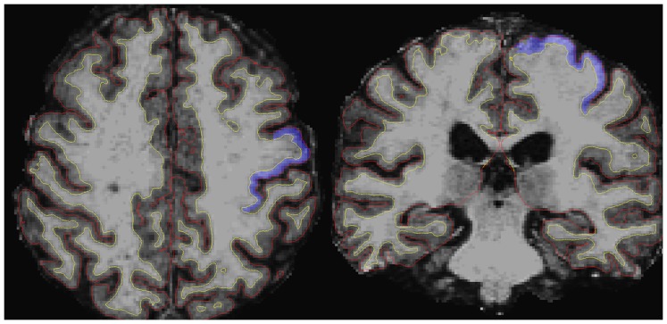
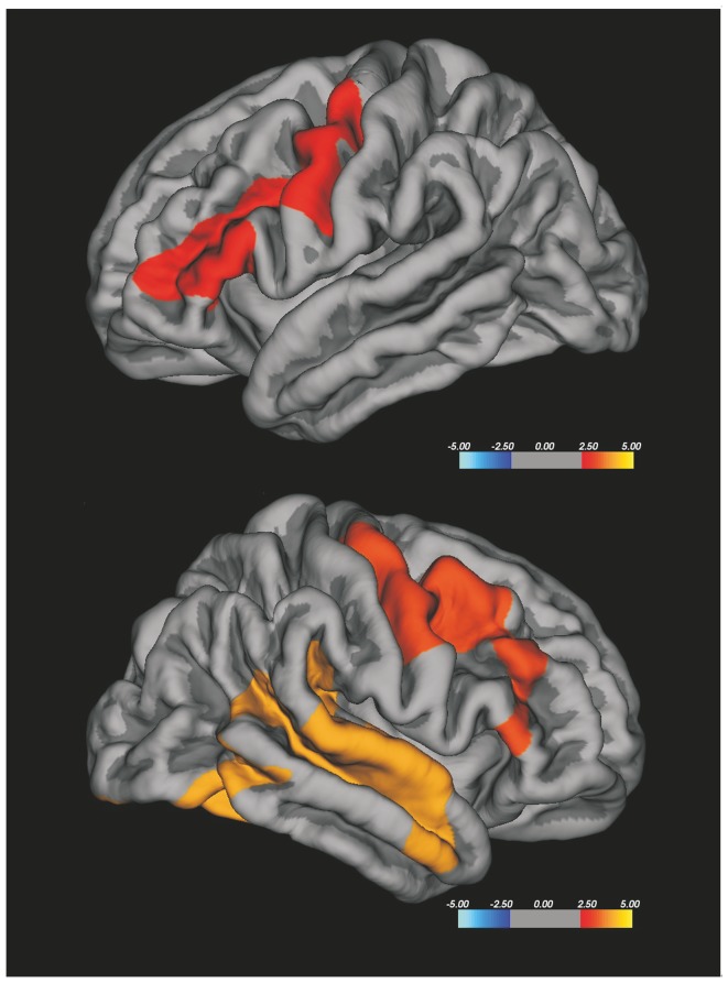
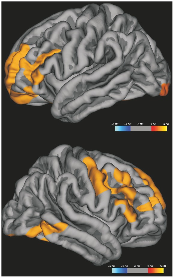
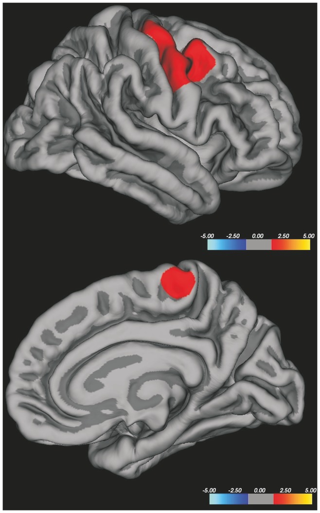
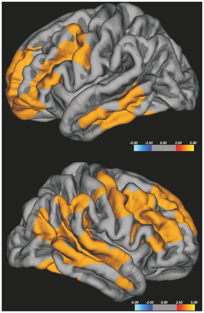
Amyotrophic lateral sclerosis (ALS) has heterogeneous clinical features that could be translated into specific patterns of brain atrophy. In the current study we have evaluated the relationship between different clinical expressions of classical ALS and measurements of brain cortical thickness. Cortical thickness analysis was conducted from 3D-MRI using FreeSurfer software in 29 ALS patients and 20 healthy controls. We explored three clinical traits of the disease, subdividing the patients into two groups for each of them: the bulbar or spinal onset, the higher or lower upper motor neuron burden, the faster or slower disease progression. We used both a whole brain vertex-wise analysis and a ROI analysis on primary motor areas. ALS patients showed cortical thinning in bilateral precentral gyrus, bilateral middle frontal gyrus, right superior temporal gyrus and right occipital cortex. ALS patients with higher upper motor neuron burden showed a significant cortical thinning in the right precentral gyrus and in other frontal extra-motor areas, compared to healthy controls. ALS patients with spinal onset showed a significant cortical thinning in the right precentral gyrus and paracentral lobule, compared to healthy controls. ALS patients with faster progressive disease showed a significant cortical thinning in widespread bilateral frontal and temporal areas, including the bilateral precentral gyrus, compared to healthy controls. Focusing on the primary motor areas, the ROI analysis revealed that the mean cortical thickness values were significantly reduced in ALS patients with higher upper motor neuron burden, spinal onset and faster disease progression related to healthy controls. In conclusion, the thickness of primary motor cortex could be a useful surrogate marker of upper motor neuron involvement in ALS; also our results suggest that cortical thinning in motor and non motor areas seem to reflect the clinical heterogeneity of the disease.


Conflict of interest statement

Competing Interests: Dr Simone received honoraria from Genzyme Sanofi-Aventis, TEVA, Bayer, and Biogen for educational lectures. Dr Logroscino received honoraria from Pfeizer and Lilly Pharmaceutical, Novartis, and Lundbeck for educational lectures. This does not alter the authors' adherence to all the PLoS ONE policies on sharing data and materials.

Figures

Figure 1
Figure 1. Example of the CTh boundaries of an ALS patients.
Axial and coronal T1-weighted images of an ALS patient. White matter surface (yellow line) and pial surface (red line), which are the boundaries of cerebral cortex, have been automatically obtained by Freesurfer. The primary motor area (blu) has been automatically labeled according to the Desikan/Killiany Atlas.
Figure 2
Figure 2. Vertex-wise analysis of CTh in ALS patients related to HC.
ALS patients revealed cortical thinning in bilateral precentral cortex, bilateral middle frontal gyrus, right superior temporal gyrus and right occipital cortex. The colour bar scale represents t values.
Figure 3
Figure 3. Vertex-wise analysis of CTh in ALS patients with higher UMN burden compared to HC.
ALS patients with higher UMN burden showed cortical thinning in the right precentral gyrus, in bilateral superior, middle and inferior frontal gyri, in the right inferior temporal gyrus and in left lateral occipital cortex. The colour bar scale represents t values.
Figure 4
Figure 4. Vertex-wise analysis of CTh in ALS patients with spinal onset related to HC.
Spinal onset ALS patients showed a significant thinning in right precentral and paracentral gyri; no differences were revealed in the left hemisphere. The colour bar scale represents t values.
Figure 5
Figure 5. Vertex-wise analysis of CTh in ALS patients with faster progression compared to HC.
Faster progressing ALS patients showed relevant thinning in widespread bilateral frontal and temporal areas, including the bilateral precentral gyrus. The colour bar scale represents t values.

Similar articles

Cited by

References

    1. Mitchell JD, Borasio GD (2007) Amyotrophic lateral sclerosis. Lancet 369: 2031–2041. - PubMed
    1. Agosta F, Chiò A, Cosottini M, De Stefano N, Falini A, et al. (2010) The present and the future of neuroimaging in amyotrophic lateral sclerosis. AJNR Am J Neuroradiol 31: 1769–1777. - PMC - PubMed
    1. Turner MR, Modo M (2010) Advances in the application of MRI to amyotrophic lateral sclerosis. Expert Opin Med Diagn 4: 483–496. - PMC - PubMed
    1. Strong MJ (2001) Progress in clinical neurosciences: the evidence for ALS as a multisystems disorder of limited phenotypic expression. Can J Neurol Sci 28: 283–298. - PubMed
    1. Mezzapesa DM, Ceccarelli A, Dicuonzo F, Carella A, De Caro MF, et al. (2007) Whole-brain and regional brain atrophy in amyotrophic laterals clerosis. AJNR Am J Neuroradiol 28: 255–259. - PMC - PubMed

Publication types