

Stem cell antigen 1-positive mesenchymal cells are the origin of follicular cells during thyroid regeneration
- PMID: 24278321
- PMCID: PMC3836768
- DOI: 10.1371/journal.pone.0080801
Stem cell antigen 1-positive mesenchymal cells are the origin of follicular cells during thyroid regeneration
Abstract
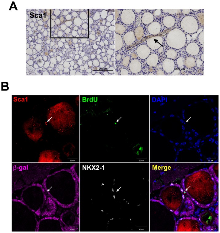
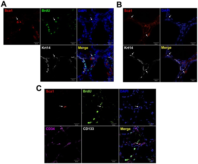
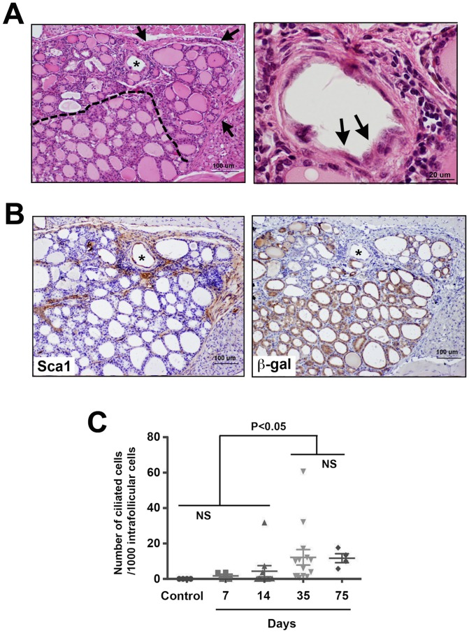
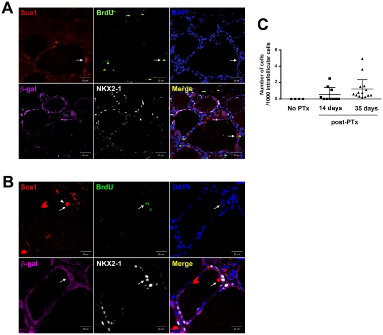
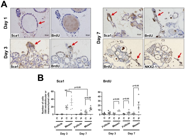
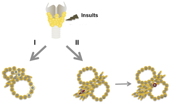
Many tissues are thought to contain adult stem/progenitor cells that are responsible for repair of the tissue where they reside upon damage and/or carcinogenesis, conditions when cellular homeostasis becomes uncontrolled. While the presence of stem/progenitor cells of the thyroid has been suggested, how these cells contribute to thyroid regeneration remains unclear. Here we show the origin of thyroid follicular cells and the process of their maturation to become follicular cells during regeneration. By using β-galactosidase (β-gal) reporter mice in conjunction with partial thyroidectomy as a model for thyroid regeneration, and bromodeoxyuridine (BrdU) long label-retaining cell analysis, we demonstrated that stem cell antigen 1 (Sca1) and BrdU-positive, but β-gal and NKX2-1 negative cells were found in the non-follicular mesenchymal area 7 days after partial thyroidectomy. They temporarily co-expressed cytokeratin 14, and were observed in part of follicles by day 35 post-partial thyroidectomy. Sca1, BrdU, β-gal, and NKX2-1-positive cells were found 120 days post-partial thyroidectomy. These results suggested that Sca1 and BrdU positive cells may participate in the formation of new thyroid follicles after partial thyroidectomy. The process of thyroid follicular cell regeneration was recapitulated in ex vivo thyroid slice collagen gel culture studies. These studies will facilitate research on thyroid stem/progenitor cells and their roles in thyroid diseases, particularly thyroid carcinomas.
Conflict of interest statement
Figures
Similar articles
-
An Adult Mouse Thyroid Side Population Cell Line that Exhibits Enriched Epithelial-Mesenchymal Transition.Thyroid. 2017 Mar;27(3):460-474. doi: 10.1089/thy.2016.0130. Epub 2017 Feb 17. Thyroid. 2017. PMID: 28125936 Free PMC article.
-
Thyroid regeneration: characterization of clear cells after partial thyroidectomy.Endocrinology. 2012 May;153(5):2514-25. doi: 10.1210/en.2011-1365. Epub 2012 Mar 27. Endocrinology. 2012. PMID: 22454152 Free PMC article.
-
Regeneration of thyroid follicles from primordial cells in a murine thyroidectomized model.Lab Invest. 2017 Apr;97(4):478-489. doi: 10.1038/labinvest.2016.158. Epub 2017 Jan 23. Lab Invest. 2017. PMID: 28112758 Free PMC article.
-
Thyroid regeneration: how stem cells play a role?Front Endocrinol (Lausanne). 2014 Apr 14;5:55. doi: 10.3389/fendo.2014.00055. eCollection 2014. Front Endocrinol (Lausanne). 2014. PMID: 24782833 Free PMC article. Review.
-
Commonly used mesenchymal stem cell markers and tracking labels: Limitations and challenges.Histol Histopathol. 2013 Sep;28(9):1109-16. doi: 10.14670/HH-28.1109. Epub 2013 Apr 16. Histol Histopathol. 2013. PMID: 23588700 Free PMC article. Review.
Cited by
-
Progress Toward and Challenges Remaining for Thyroid Tissue Regeneration.Endocrinology. 2023 Aug 28;164(10):bqad136. doi: 10.1210/endocr/bqad136. Endocrinology. 2023. PMID: 37690118 Free PMC article. Review.
-
An Adult Mouse Thyroid Side Population Cell Line that Exhibits Enriched Epithelial-Mesenchymal Transition.Thyroid. 2017 Mar;27(3):460-474. doi: 10.1089/thy.2016.0130. Epub 2017 Feb 17. Thyroid. 2017. PMID: 28125936 Free PMC article.
-
Normal vs cancer thyroid stem cells: the road to transformation.Oncogene. 2016 Feb 18;35(7):805-15. doi: 10.1038/onc.2015.138. Epub 2015 May 11. Oncogene. 2016. PMID: 25961919 Review.
-
Engineering a functional thyroid as a potential therapeutic substitute for hypothyroidism treatment: A systematic review.Front Endocrinol (Lausanne). 2022 Dec 2;13:1065410. doi: 10.3389/fendo.2022.1065410. eCollection 2022. Front Endocrinol (Lausanne). 2022. PMID: 36531472 Free PMC article.
-
Stem Cell Therapy for Thyroid Diseases: Progress and Challenges.Curr Ther Res Clin Exp. 2022 Mar 5;96:100665. doi: 10.1016/j.curtheres.2022.100665. eCollection 2022. Curr Ther Res Clin Exp. 2022. PMID: 35371349 Free PMC article. Review.
References
-
- Thomas T, Nowka K, Lan L, Derwahl M (2006) Expression of endoderm stem cell markers: evidence for the presence of adult stem cells in human thyroid glands. Thyroid 16: 537–544. - PubMed
-
- Lan L, Cui D, Nowka K, Derwahl M (2007) Stem cells derived from goiters in adults form spheres in response to intense growth stimulation and require thyrotropin for differentiation into thyrocytes. J Clin Endocrinol Metab 92: 3681–3688. - PubMed
-
- Fierabracci A, Puglisi MA, Giuliani L, Mattarocci S, Gallinella-Muzi M (2008) Identification of an adult stem/progenitor cell-like population in the human thyroid. J Endocrinol 198: 471–487. - PubMed
Publication types
MeSH terms
Substances
Grants and funding
LinkOut - more resources
Full Text Sources
Other Literature Sources
