. 2013 Nov 20;8(11):e80809.
doi: 10.1371/journal.pone.0080809. eCollection 2013.

Loss of the cytoskeletal protein Pdlim7 predisposes mice to heart defects and hemostatic dysfunction

Affiliations

Loss of the cytoskeletal protein Pdlim7 predisposes mice to heart defects and hemostatic dysfunction

Jennifer Krcmery et al. PLoS One. .

Abstract

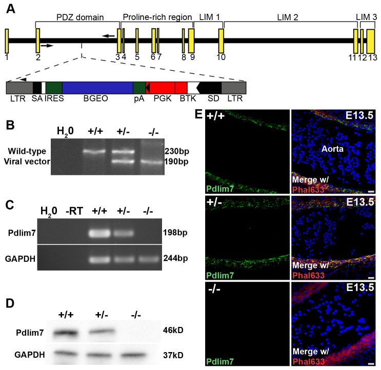
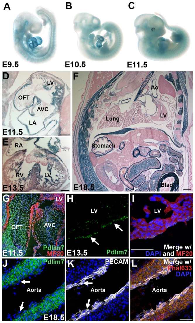
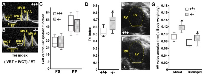
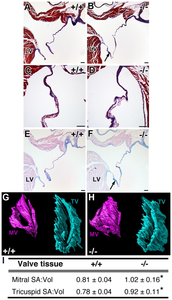
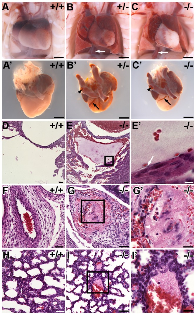
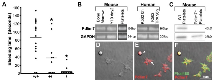
The actin-associated protein Pdlim7 is essential for heart and fin development in zebrafish; however, the expression and function of this PDZ-LIM family member in the mammal has remained unclear. Here, we show that Pdlim7 predominantly localizes to actin-rich structures in mice including the heart, vascular smooth muscle, and platelets. To test the requirement for Pdlim7 in mammalian development and function, we analyzed a mouse strain with global genetic inactivation of Pdlim7. We demonstrate that Pdlim7 loss-of-function leads to significant postnatal mortality. Inactivation of Pdlim7 does not disrupt cardiac development, but causes mild cardiac dysfunction in adult mice. Adult Pdlim7(-/-) mice displayed increased mitral and tricuspid valve annulus to body weight ratios. These structural aberrations in Pdlim7(-/-) mice were supported by three-dimensional reconstructions of adult cardiac valves, which revealed increased surface area to volume ratios for the mitral and tricuspid valve leaflets. Unexpectedly, we found that loss of Pdlim7 triggers systemic venous and arterial thrombosis, leading to significant mortality shortly after birth in Pdlim7(+/-) (11/60) and Pdlim7(-/-) (19/35) mice. In line with a prothrombotic phenotype, adult Pdlim7(-/-) mice exhibit dramatically decreased tail bleed times compared to controls. These findings reveal a novel and unexpected function for Pdlim7 in maintaining proper hemostasis in neonatal and adult mice.


Conflict of interest statement

Competing Interests: The authors have declared that no competing interests exist.

Figures

Figure 1
Figure 1. Generation of Pdlim7 mutant mice.
Schematic representation of the retroviral integration site within intron two of the Pdlim7 gene (A). Exons are depicted by yellow boxes with WT and viral-specific genotyping primers represented by arrows and an arrowhead, respectively. Genotyping reveals a 230bp PCR fragment representing the WT allele and a 190bp product for the viral integration (B). Semi-quantitative RT-PCR demonstrates absence of Pdlim7 gene transcripts in adult uteri of Pdlim7-/- mice using GAPDH as control (C). Western blot shows that Pdlim7-/- and Pdlim7+/- mice express only -1.18 ± 2.64% and 29.36 ± 14.92% Pdlim7 protein compared to WT controls, respectively (D). Sagittal section through the aorta demonstrates Pdlim7 antibodies (green) localize to filamentous actin (red) of smooth muscle in WT, but not Pdlim7-/- embryos (E), further verifying loss of Pdlim7 protein in null mice (control DAPI nuclei, blue). LTR = long terminal repeat; SA and SD = splice acceptor and donor sites, respectively; IRES = internal ribosomal entry site; BGEO = lacZ gene; pA = polyadenylation signal; PGK = phosphoglycerate kinase gene; BTK = Bruton’s tyrosine kinase gene.
Figure 2
Figure 2. Pdlim7 is dynamically expressed in actin-rich structures throughout murine development.
β-galactosidase staining of whole mount embryos identifies Pdlim7 early in the developing heart, forelimbs, hindlimbs, and somites (A-C). In E11.5 and E13.5 heart sections, β-galactosidase staining is localized to the trabeculated regions of the ventricles, atrial walls, and interventricular and atrial septa (D-E); of note: the β-galactosidase reaction time chosen does not reveal the lower levels of Pdlim7 expression in the cardiac cushion or valve cells. Sagittal sections of E18.5 embryos show Pdlim7 expression throughout the smooth muscle, including the lung alveoli, stomach, aorta, and bladder (F). Immunostaining of E11.5 hearts demonstrates Pdlim7 (green) localization to the outflow tract and atrioventricular cushions with MF20 positive myocardial cells (red) (G). Pdlim7 (green) proteins are also detected in the epicardium of E13.5 embryos (arrows, H-I) with MF20 positive myocardial cells (red) and DAPI nuclei (blue). In the aorta, Pdlim7 proteins (green) localize to filamentous actin (red) of smooth muscle, but not PECAM-positive endothelium (white) (arrows, J-L), control DAPI nuclei (blue). Scale bar = 200μm (D-E) and 50 μm (F, I, L). Ao = Aorta; AVC = atrioventricular cushion; Blad = bladder; LA = left atrium; LV = left ventricle; OFT = outflow tract; RA = right atrium; RV = right ventricle.
Figure 3
Figure 3. Adult Pdlim7-/- mice exhibit mild cardiac dysfunction with increased valve annulus dimensions.
Representative Doppler profile in the apical-4 chamber view of mitral valve inflow velocities (A-B) and mitral valve annulus measurement (E-F) from WT (n=8) and Pdlim7-/- (n=9) mice. Box-and-whisker plots reveal normal fractional shortening (FS) and ejection fraction (EF) in Pdlim7-/- mice (C), but an increased Tei index (D) and increased mitral and tricuspid annulus dimensions to body weight ratios (G) as compared to controls, *p<0.01. A = late filling; AV = atrioventricular; E = early filling; ET = ejection time; IVCT = isovolumetric contraction time; IVRT = isovolumetric relaxation time; LV = left ventricle; MV = mitral valve; RV = right ventricle.
Figure 4
Figure 4. Atrioventricular valves of adult Pdlim7-/- mice display abnormal morphology.
Histological sections from 3-month old WT and Pdlim7-/- hearts stained with Masson’s Trichrome (A-D) and Alcian blue (E-F) exhibit normal distribution of collagens (blue, A-D) and glycosaminoglycans (blue, E-F), but elongated mitral valves in Pdlim7-/- mice (arrows, B, F; n=5) compared to WT controls (A, E; n=3). Scale bar = 100 μm. Representative 3D reconstructions of serial sections of mitral (pink) and tricuspid (turquoise) valves from a WT (G) and Pdlim7-/- (H) mouse in the same anterodorsolateral perspective. Table providing quantification of increased surface area to volume ratios of atrioventricular valves in Pdlim7-/- (mitral, n=5; tricuspid, n=4) compared to WT (mitral, n=6; tricuspid, n=5) controls (I). *p<0.04. LV = left ventricle; MV = mitral valve; SA = surface area; TV = tricuspid valve; Vol = volume.
Figure 5
Figure 5. Pathological analysis of non-surviving Pdlim7 mutant pups reveals pre-mortem thrombi.
Pdlim7 +/- (n=3) and Pdlim7 -/- (n=11) perinatal lethal pups exhibit extensive blood clots in the heart, arteries (arrowhead), and veins (arrows, B-C’) associated with atrial dilation and lung congestion compared to WT (n=7) controls (A-A’). Example of pre-mortem blood clots in the right ventricle (E-E’), umbilical vessel (G-G’), and lung alveoli (I-I’) of a Pdlim7-/- perinatal lethal pup compared to WT control (D, F, H). Boxes depict location of high magnification image in E’, G’, and I’. Arrows point to attachment of the thrombus to the vessel wall (E’, I’) and arrowhead points to recanalization of the clot (G’). Scale bar = 1mm (A-C’), 50 µm (D-I), 20 µm (G’, I’), and 10µm (E’).
Figure 6
Figure 6. Pdlim7 is expressed in platelets and adult Pdlim7 deficient mice display hemostatic dysfunction.
Upon tail clip, blood from 3-week old Pdlim7-/- (n=9) mice clotted immediately compared to WT (n=16) controls with Pdlim7+/- mice (n=10) exhibiting an intermediate phenotype (A). Horizontal line denotes mean bleed time for each genotype *p<0.01. Semi-quantitative RT-PCR identifies Pdlim7 gene transcripts in WT mouse bone marrow, megakaryocyte cell line Y10 (undifferentiated, day2), and platelets as well as human bone marrow cell line K562 (undifferentiated, 0h; differentiated into proplatelets using TPA for 96h) using GAPDH as a control (B). Western blot analysis of platelets from WT mice demonstrates the presence of Pdlim7 proteins and the absence of Pdlim7 proteins in Pdlim7-/- mice using GAPDH as a loading control (C). Confocal images of WT platelets spread on glass and immunostained with anti-Pdlim7 antibodies (red) and counterstained for F-actin (green) (D-F). Notably, Pdlim7 is distributed in resting (arrowhead) and spreading platelets (arrow) (E). Scale bar = 5µm.

Similar articles

Cited by

References

    1. Krcmery J, Camarata T, Kulisz A, Simon HG (2010) Nucleocytoplasmic functions of the PDZ-LIM protein family: new insights into organ development. Bioessays 32: 100-108. doi:10.1002/bies.200900148. PubMed: 20091751. - DOI - PMC - PubMed
    1. te Velthuis AJ, Bagowski CP (2007) PDZ and LIM domain-encoding genes: molecular interactions and their role in development. ScientificWorldJournal 7: 1470-1492. doi:10.1100/tsw.2007.232. PubMed: 17767364. - DOI - PMC - PubMed
    1. Camarata T, Snyder D, Schwend T, Klosowiak J, Holtrup B et al. (2010b) Pdlim7 is required for maintenance of the mesenchymal/epidermal Fgf signaling feedback loop during zebrafish pectoral fin development. BMC Dev Biol 10: 104. doi:10.1186/1471-213X-10-104. PubMed: 20950450. - DOI - PMC - PubMed
    1. Pashmforoush M, Pomiès P, Peterson KL, Kubalak S, Ross J Jr. et al. (2001) Adult mice deficient in actinin-associated LIM-domain protein reveal a developmental pathway for right ventricular cardiomyopathy. Nat Med 7: 591-597. doi:10.1038/87920. PubMed: 11329061. - DOI - PubMed
    1. Zhou Q, Chu PH, Huang C, Cheng CF, Martone ME, et al. (2001) Ablation of Cypher, a PDZ-LIM domain Z-line protein, causes a severe form of congenital myopathy. J Cell Biol 155: 605-612 - PMC - PubMed

Publication types

MeSH terms