. 2013 Nov 22;8(11):e80849.
doi: 10.1371/journal.pone.0080849. eCollection 2013.

HDAC4 does not act as a protein deacetylase in the postnatal murine brain in vivo

Affiliations

HDAC4 does not act as a protein deacetylase in the postnatal murine brain in vivo

Michal Mielcarek et al. PLoS One. .

Abstract

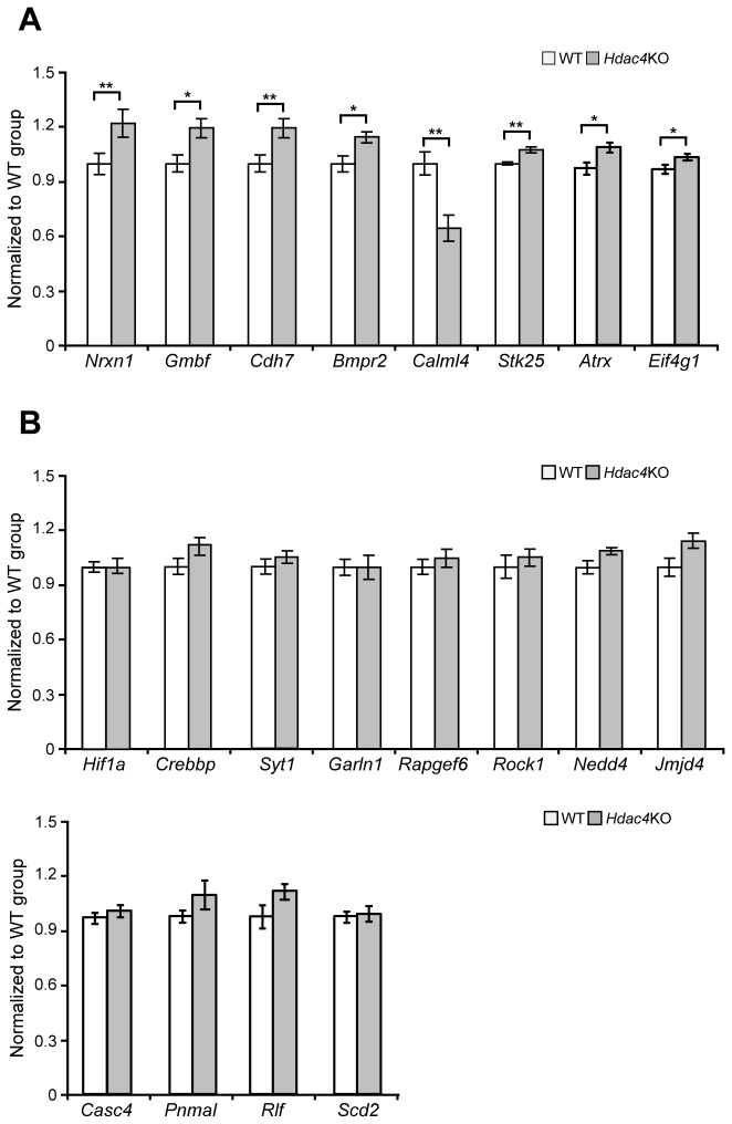
Reversible protein acetylation provides a central mechanism for controlling gene expression and cellular signaling events. It is governed by the antagonistic commitment of two enzymes families: the histone acetyltransferases (HATs) and the histone deacetylases (HDACs). HDAC4, like its class IIa counterparts, is a potent transcriptional repressor through interactions with tissue specific transcription factors via its N-terminal domain. Whilst the lysine deacetylase activity of the class IIa HDACs is much less potent than that of the class I enzymes, HDAC4 has been reported to influence protein deacetylation through its interaction with HDAC3. To investigate the influence of HDAC4 on protein acetylation we employed the immunoaffinity-based AcetylScan proteomic method. We identified many proteins known to be modified by acetylation, but found that the absence of HDAC4 had no effect on the acetylation profile of the murine neonate brain. This is consistent with the biochemical data suggesting that HDAC4 may not function as a lysine deacetylase, but these in vivo data do not support the previous report showing that the enzymatic activity of HDAC3 might be modified by its interaction with HDAC4. To complement this work, we used Affymetrix arrays to investigate the effect of HDAC4 knock-out on the transcriptional profile of the postnatal murine brain. There was no effect on global transcription, consistent with the absence of a differential histone acetylation profile. Validation of the array data by Taq-man qPCR indicated that only protamine 1 and Igfbp6 mRNA levels were increased by more than one-fold and only Calml4 was decreased. The lack of a major effect on the transcriptional profile is consistent with the cytoplasmic location of HDAC4 in the P3 murine brain.


Conflict of interest statement

Competing Interests: The authors have read the journal's policy and have the following conflicts: Cell Signaling Technologies conducted the research through a fee-for-service agreement for King’s College London. Vahri Beaumont is employed by CHDI Management Inc. to provide consulting services to CHDI Foundation. MPS and JCS are employees of Cell Signaling Technologies. There are no patents, products in development or marketed products to declare. This does not alter the authors’ adherence to all the PLoS ONE policies on sharing data and materials, as detailed in the online guide for authors.

Figures

Figure 1
Figure 1. Absence of HDAC4 causes only minor changes to the neonate brain acetylome.
(A) Histogram showing normalized log2 ratios between HDAC4 KO and WT brains for all peptides identified. The VEGF peptide with a >2.5-fold change is highlighted. (B) Relative abundance for VEGF K324, 326, 329 acetylated peptide across all samples (control and HDAC4 KO pool1 and pool 2).
Figure 2
Figure 2. Validation of the genes predicted to be upregulated in Hdac4 knock-out P3 brains.
(A) Prm1 (protamine 1) and Igbp6 (insulin-growth factor binding protein 6) transcript levels were significantly upregulated in Hdac4KO P3 brains by 14-fold and 3-fold respectively. (B) Synpo (Synaptopodin) was modestly upregulated in Hdac4KO P3 brains whereas Sytl2 (synaptotagmin like 2 protein) was unchanged. (C) Runx2 (runt-related transcription factor 2), Ngef (neuronal guanine nucleotide exchange factor), Jmjd1C (jumonji domain containing 1C), Hsbp3 (heat shock protein 3), Armc9 (armadilo repeat containing protein 9), Prkcc (protein kinase C gamma), Pam (peptidylglycine alpha-amidating monooxygenase), CamkIV (calcium /calmodulin-dependent protein kinase IV), Ramp1 (receptor (calcitonin) activity modifying protein 1), Retnlα (resistin like alpha protein), Slc26α8 (solute carrier family 26, member 8) were modestly upregulated in Hdac4KO P3 brains whereas Parp11 (poly(ADP-ribose) polymerase member 11), Rcor3 (REST corepresor 3), Ing5 (inhibitor of growth family 5) Nrp2 (neuropilin 2) and Farsb (phenylalanyl-tRNA synthetase, beta subunit) were unchanged. All mRNA expression levels were assessed by Taqman qPCR and presented as a relative expression ratio to the geometric mean of three housekeeping genes Atp5b, Canx, Rpl13α. Error bars are S.E.M (n>7). *p<0.05, **p<0.01, ***p<0.001.
Figure 3
Figure 3. Validation of the genes predicted to be downregulated in Hdac4 knock-out P3 brains.
(A) Nrxn1 (neurexin1), Gmbf (glia maturation factor beta), Cdh7 (cadherin 7), Bmpr2 (bone morphogenic protein receptor, type II), Stk25 (serine/threonine kinase 25), Atrx (alpha thalassemia/mental retardation syndrome X-linked homolog) and Eif4g1 (eukaryotic translation initiation factor 4, gamma 1) were modestly upregulated in Hdac4KO P3 brains. Calml4 (calmodulin-like 4) was the only gene that was downregulated (B) The expression level of Hif1α (hypoxia inducible factor 1 alpha subunit), Crebbp (CREB-binding protein), Syt1 (synaptotagemin 1) Garln1 (GTPase activating RANGAP domain like 1 protein), Rapgef6 (Rap guanine nucleotide exchange factor GEF6), Rock1 (Rho-associated coiled-coil containing protein kinase 1), Nedd4 (NEDD4 binding protein), Jmjd4 (jumonji containing protein 4), Casc4 (cancer susceptibility candidate 4), Pnmal (PNMA-like 2), Rlf (rearranged L-myc fusion sequence) and Scd2 (stearoyl-Coenzyme A desaturase 2) did not change between Hdac4KO and WT P3 brains. All mRNA expression levels were assessed by Taqman qPCR and presented as a relative expression ratio to the geometric mean of three housekeeping genes Atp5b, Canx, Rpl13α. Error bars are S.E.M (n>7). *p<0.05, **p<0.01.
Figure 4
Figure 4. HDAC4 is localised to the cytoplasm in the P3 brain.
(A) Western blot showing that HDAC4 is localised exclusively to the cytoplasm and absent in Hdac4KO brains. (B) Confocal images demonstrating that HDAC4 is localised to the cytoplasm. Scale bar = 25 μm.

References

    1. Butler R, Bates GP (2006) Histone deacetylase inhibitors as therapeutics for polyglutamine disorders. Nat Rev Neurosci 7: 784-796. doi:10.1038/nrn1989. PubMed: 16988654. - DOI - PubMed
    1. Verdin E, Dequiedt F, Kasler HG (2003) Class II histone deacetylases: versatile regulators. Trends Genet 19: 286-293. doi:10.1016/S0168-9525(03)00073-8. PubMed: 12711221. - DOI - PubMed
    1. Parra M, Verdin E (2010) Regulatory signal transduction pathways for class IIa histone deacetylases. Curr Opin Pharmacol 10: 454-460. doi:10.1016/j.coph.2010.04.004. PubMed: 20447866. - DOI - PubMed
    1. Chawla S, Vanhoutte P, Arnold FJL, Huang CLH, Bading H (2003) Neuronal activity-dependent nucleocytoplasmic shuttling of HDAC4 and HDAC5. J Neurochem 85: 151-159. doi:10.1046/j.1471-4159.2003.01648.x. PubMed: 12641737. - DOI - PubMed
    1. McKinsey TA, Zhang CL, Olson EN (2000) Activation of the myocyte enhancer factor-2 transcription factor by calcium/calmodulin-dependent protein kinase-stimulated binding of 14-3-3 to histone deacetylase 5. Proc Natl Acad Sci U S A 97: 14400-14405. doi:10.1073/pnas.260501497. PubMed: 11114197. - DOI - PMC - PubMed

Publication types