. 2013 Nov 20;8(11):e81019.
doi: 10.1371/journal.pone.0081019. eCollection 2013.

The Nogo-B-PirB axis controls macrophage-mediated vascular remodeling

Affiliations

The Nogo-B-PirB axis controls macrophage-mediated vascular remodeling

Yuka Kondo et al. PLoS One. .

Abstract

Objective: Nogo-B mediates vascular protection and facilitates monocyte- and macrophage-dependent vascular remodeling. PirB is an alternate receptor for Nogo-B, but a role for the Nogo-PirB axis within the vascular system has not been previously reported. We examined whether Nogo-B or PirB play a role in regulating macrophage-mediated vascular remodeling and hypothesized that endothelial Nogo-B regulates vein graft macrophage infiltration via its alternate receptor PirB.

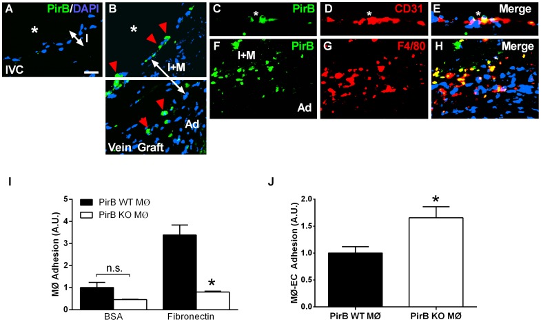
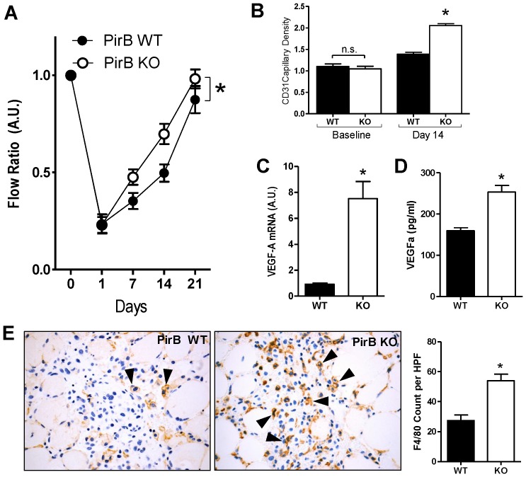
Methods: Vein grafts were performed using Nogo and PirB wild type and knockout mice. Human vein grafts were similarly analyzed. The hindlimb ischemia model was performed in PirB wild type and knockout mice. Accompanying in vitro work included isolation of macrophages from PirB wild type and knockout mice.

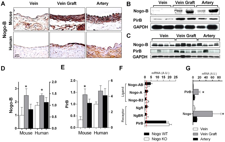
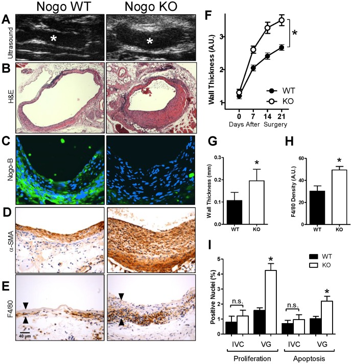
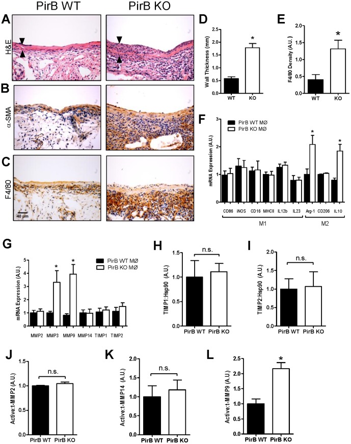
Results: Increased Nogo-B and PirB mRNA transcripts and protein expression were observed within mouse and human vein grafts. Both Nogo knockout and PirB knockout vein grafts showed increased wall thickness and increased numbers of F4/80-positive macrophages. Macrophages derived from PirB knockout mice had increased adhesion to fibronectin, increased EC-specific binding, and increased numbers of mRNA transcripts of M2 markers as well as MMP3 and MMP9. PirB knockout vein grafts had increased active MMP9 compared to wild type vein grafts. PirB knockout mice had increased recovery from hindlimb ischemia and increased macrophage infiltration compared to wild type mice.

Conclusions: Vein graft adaptation shows increased expression of both Nogo-B and PirB. Loss of PirB, or its endothelial ligand Nogo-B, results in increased inflammatory cell infiltration and vein graft wall thickening. These findings suggest that PirB regulates macrophage activity in vein grafts and that Nogo-B in the vein graft limits macrophage infiltration and vein graft thickening. PirB may play a more general role in regulating macrophage responses to vascular injury. Macrophage inhibition via Nogo-PirB interactions may be an important mechanism regulating vein graft adaptation to the arterial circulation.


Conflict of interest statement

Competing Interests: The authors have declared that no competing interests exist.

Figures

Figure 1
Figure 1. Increased Nogo-B and PirB expression in mouse and human vein grafts.
(A) Representative immunohistochemical staining images in mouse (top row) and human (bottom row) vein (left), vein graft (center), and aorta (right). n = 4. Scale bar, 40 µm. (B) Representative Western blot analysis showing Nogo-B and PirB up-regulation in individual mouse vein grafts; n = 2 matched specimens are shown of n = 4 samples. (C) Representative Western blot analysis showing Nogo-B and PirB up-regulation in individual human vein grafts; n = 4 unmatched specimens are shown. (D) Bar graph shows densitometry of Nogo-B bands shown in Panels (B) and (C) during mouse and human vein graft adaptation. *, p<0.05; t-test (vein vs. vein graft). (E) Bar graph shows densitometry of PirB bands shown in Panels (B) and (C) during mouse and human vein graft adaptation. *, p<0.05; t-test (vein vs. vein graft). (F) Bar graph shows summary of gene expression during mouse vein graft adaptation; the number of mRNA transcripts expressed during vein graft adaptation is compared to the number expressed in the wild type pre-implantation inferior vena cava (IVC), denoted by the red broken line. n = 4. ▪, Nogo WT; □, Nogo KO vein grafts. (G) Bar graph shows Nogo-B and PirB mRNA transcripts during human vein graft adaptation. Analysis of n = 8 veins, 6 vein grafts, and 5 arterial human specimens. *, p<0.05; t-test (vein vs. vein graft).
Figure 2
Figure 2. PirB mediates adhesion in vitro and in vivo.
(A) Representative immunofluorescence image showing lack of PirB signal in the pre-implantation vein. IVC, inferior vena cava. Scale bar, 20 µm. (B) PirB positive cells localized to the luminal surface and the interface between the medial and adventitial layers. *, vessel lumen; red arrowhead, PirB positive cells; I, intimal layer; I+M, intima-medial layer; Ad, adventitial layer. (C–E) PirB-positive cells on the vein graft luminal vessel surface (C) did not co-localize with CD31-positive endothelial cells (E). *, vessel lumen; white arrows, PirB-positive cells. (F–H) PirB-positive cells in between the medial and adventitial layers (F) co-localize with F4/80-positive cells (G,H). I+M, intima-medial layer; Ad, adventitial layer. n = 4. (I) Bar graph shows macrophage adhesion to bovine serum albumin (BSA) or fibronectin. Macrophages were derived from PirB WT (▪) or PirB KO (□) mice. n.s., not significant. n = 8. *, p<0.0001; ANOVA; post-hoc testing p<0.05. (J) Bar graph shows macrophage adhesion to endothelial cells. Macrophages were derived from PirB WT (▪) or PirB KO (□) mice and activated with TNF-α. *, p = 0.0499, t-test; n = 3.
Figure 3
Figure 3. Nogo-B limits F4/80-positive cell infiltration during vein graft adaptation.
(A) Representative ultrasound images of mouse vein grafts. Nogo KO grafts (right panel) had significantly thicker graft walls compared to those of Nogo WT grafts (left panel). *, lumen. (B) Representative H&E image of WT and Nogo KO mouse vein grafts. Scale bar, 100 µm. (C) Representative immunofluorescence images of Nogo staining in WT and KO mouse vein grafts; no staining is present in Nogo KO vein grafts. Scale bar, 50 µm. (D) Representative image of smooth muscle alpha-actin (SMA) staining of Nogo WT and KO mouse vein grafts. (E) Representative images of Nogo WT and KO vein grafts with immunohistochemical staining for F4/80. Arrowheads show vein graft wall. n = 20, 25. (F) Line graph shows the time course of vein graft wall thickening as determined by ultrasound imaging. Nogo KO grafts had approximately 40 percent thicker walls compared to those of wild type grafts. *, p = 0.0041; ANOVA. (G) Bar graph shows summary of morphological analysis of wall thickness of Nogo WT and KO grafts. *, p = 0.0061; t-test; n = 5. (H) Bar graph shows summary of densitometry of F4/80 staining. *, p = 0.0011; t-test. n = 20, 25. (I) Bar graphs show summary of proliferation and apoptosis indices in mouse vein grafts. *, p<0.0001, ANOVA; post-hoc testing p<0.05; n = 8, 16, 8, 32 in each of the 4 groups, respectively.
Figure 4
Figure 4. PirB limits F4/80-positive cell infiltration during vein graft adaptation.
(A) Representative H&E image of PirB WT and KO mouse vein grafts. Scale bar, 100 µm. Arrowheads show vein graft wall. n = 7. (B) Representative image of smooth muscle alpha-actin (SMA) staining of PirB WT and KO mouse vein grafts. n = 4. (C) Representative images of PirB WT and KO vein grafts with immunohistochemical staining for F4/80. n = 4. (D) Bar graph shows summary of morphological analysis of wall thickness of PirB WT and KO grafts. *, p<0.0001, t-test; n = 7. (E) Bar graph shows summary of densitometry of F4/80 staining. *, p = 0.0211, t-test; n = 4. (F) Bar graph shows summary of mRNA transcript expression in macrophages derived from PirB WT (▪) or PirB KO (□) mice. *, p<0.02, t-test. n = 6. (G) Bar graph shows summary of mRNA transcript expression in macrophages derived from PirB WT (▪) or PirB KO (□) mice. *, p<0.03, t-test. n = 6. (H) Bar graph shows mean densitometry of TIMP1 immunoreactivity in vein grafts derived from PirB WT (▪) or PirB KO (□) mice. n.s., not significant; p = 0.7859, t-test; n = 3. (I) Bar graph shows mean densitometry of TIMP2 immunoreactivity in vein grafts derived from PirB WT (▪) or PirB KO (□) mice. n.s., not significant; p = 0.8941, t-test; n = 3. (J) Bar graph shows mean densitometry of MMP2 activity in vein grafts derived from PirB WT (▪) or PirB KO (□) mice. n.s., not significant; p = 0.2252, t-test; n = 3. (K) Bar graph shows mean densitometry of MMP14 activity in vein grafts derived from PirB WT (▪) or PirB KO (□) mice. n.s., not significant; p = 0.6569, t-test; n = 4. (L) Bar graph shows mean densitometry of MMP9 activity in vein grafts derived from PirB WT (▪) or PirB KO (□) mice. *, p = 0.0109, t-test; n = 3.
Figure 5
Figure 5. PirB mediates other macrophage-mediated vascular functions.
(A) Line graph shows mean deep muscle perfusion (ratio ischemic:control leg), 0–21 days after induction of ischemia in PirB WT (▪) or PirB KO (□) mice. *, p<0.0001, ANOVA; n = 10. (B) Bar graph show summary of capillary density, at baseline (p = 0.5350, t-test; n = 3) or 14 days after induction of ischemia (*, p<0.0001, t-test; n = 3) in PirB WT (▪) or PirB KO (□) mice. (C) Bar graphs show mean VEGF-A mRNA (*, p = 0.001, t-test; n = 5) 72 hours after induction of ischemia in PirB WT (▪) and PirB KO (□) mice. (D) Bar graphs show mean VEGF-A protein secretion by PirB WT (▪) and PirB KO macrophages (□) (*, p = 0.0003, t-test; n = 6). (E) Representative photomicrographs showing F4/80 staining (black arrowheads) in gastrocnemius muscle 3 days after induction of ischemia in PirB WT (▪) or PirB KO (□) mice. Right panel shows bar graph summarizing mean number of F4/80-positive cells per hpf. *, p = 0.0016, t-test; n = 3.

References

    1. Owens CD (2010) Adaptive changes in autogenous vein grafts for arterial reconstruction: clinical implications. J Vasc Surg 51: 736–746. - PMC - PubMed
    1. Acevedo L, Yu J, Erdjument-Bromage H, Miao RQ, Kim JE, Fulton D (2004) A new role for Nogo as a regulator of vascular remodeling. Nat Med 10: 382–388. - PubMed
    1. Paszkowiak JJ, Maloney SP, Kudo FA, Muto A, Teso D, Rutland RC, Westvik TS, Pimiento JM, Tellides G, Sessa WC, Dardik A (2007) Evidence supporting changes in Nogo-B levels as a marker of neointimal expansion but not adaptive arterial remodeling. Vascul Pharmacol 46: 293–301. - PMC - PubMed
    1. Kritz AB, Yu J, Wright PL, Wan S, George SJ, Halliday C (2008) In vivo modulation of Nogo-B attenuates neointima formation. Mol Ther 16: 1798–1804. - PMC - PubMed
    1. Kudo FA, Muto A, Maloney SP, Pimiento JM, Bergaya S, et al. (2007) Venous identity is lost but arterial identity is not gained during vein graft adaptation. Arterioscler Thromb Vasc Biol 27: 1562–1571. - PubMed

Publication types

LinkOut - more resources