. 2013 Nov 21;8(11):e81061.
doi: 10.1371/journal.pone.0081061. eCollection 2013.

A novel in vitro model for microvasculature reveals regulation of circumferential ECM organization by curvature

Affiliations

A novel in vitro model for microvasculature reveals regulation of circumferential ECM organization by curvature

Sebastian F Barreto-Ortiz et al. PLoS One. .

Abstract

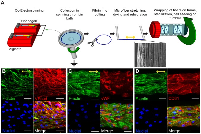
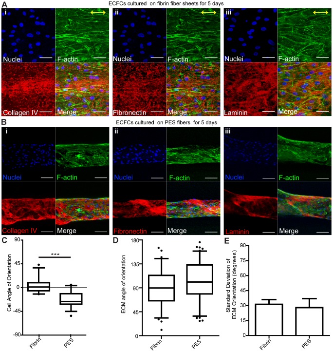
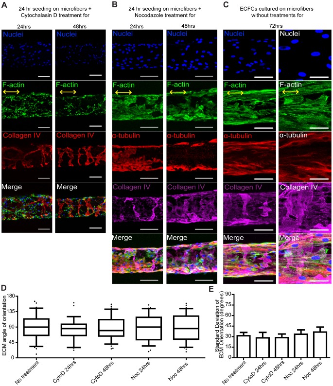
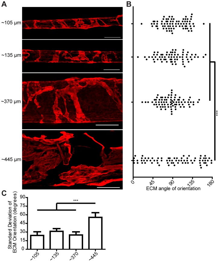
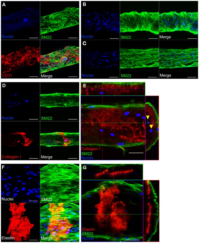
In microvascular vessels, endothelial cells are aligned longitudinally whereas several components of the extracellular matrix (ECM) are organized circumferentially. While current three-dimensional (3D) in vitro models for microvasculature have allowed the study of ECM-regulated tubulogenesis, they have limited control over topographical cues presented by the ECM and impart a barrier for the high-resolution and dynamic study of multicellular and extracellular organization. Here we exploit a 3D fibrin microfiber scaffold to develop a novel in vitro model of the microvasculature that recapitulates endothelial alignment and ECM deposition in a setting that also allows the sequential co-culture of mural cells. We show that the microfibers' nanotopography induces longitudinal adhesion and alignment of endothelial colony-forming cells (ECFCs), and that these deposit circumferentially organized ECM. We found that ECM wrapping on the microfibers is independent of ECFCs' actin and microtubule organization, but it is dependent on the curvature of the microfiber. Microfibers with smaller diameters (100-400 µm) guided circumferential ECM deposition, whereas microfibers with larger diameters (450 µm) failed to support wrapping ECM. Finally, we demonstrate that vascular smooth muscle cells attached on ECFC-seeded microfibers, depositing collagen I and elastin. Collectively, we establish a novel in vitro model for the sequential control and study of microvasculature development and reveal the unprecedented role of the endothelium in organized ECM deposition regulated by the microfiber curvature.


Conflict of interest statement

Competing Interests: The authors have declared that no competing interests exist.

Figures

Figure 1
Figure 1. ECFCs attached and aligned on fibrin hydrogel microfibers.
(A) Schematic of experimental procedure including electrospinning, ECFC seeding, and tumbling. Drawing not to scale. SEM of critical-point dried fibrin microfiber showing aligned topography on the microfiber surface. Scale bar is 10 µm. (B–D) Confocal z-stack image reconstructions of ECFCs seeded on fibrin hydrogel microfibers horizontally aligned after 5 days in culture. F-actin (phalloidin staining) is shown in green, EC-specific markers (VECad, CD31, or vWF) in red, and nuclei in blue. Yellow arrows indicate the direction of stretching and nanotopography on microfiber surface. Scale bars are 50 µm. n≥2 per stain with quadruplicates.
Figure 2
Figure 2. ECFCs deposit ECM circumferentially on fibrin hydrogel microfibers.
Confocal z-stack image reconstructions of ECFCs on fibrin microfibers after (A) 1 and (B) 5 days in culture. Scale bars are 200 µm. (C) High magnification confocal images of laminin, fibronectin and collagen IV wrapping around the fibrin microfibers. (D) TEM images of cross-sectional slices of a cell-fibrin microfiber construct after 5 days in culture (i–ii) with cells and (iii) without cells. (ii) is a higher magnification image for the boxed area in (i). F  =  Fibrin; E  =  ECM; C  =  Cells. (E) Cross-sectional projections of confocal z-stack images of ECFCs on fibrin microfibers after 5 days. F-actin (phalloidin) is shown in green, ECM proteins (collagen IV, laminin, fibronectin) in red or magenta, and nuclei in blue. Yellow arrows indicate the direction of nanotopography on microfiber surface. A n≥2, B–E n≥5 per stain with quadruplicates.
Figure 3
Figure 3. Nanotopography and geometry differently effect ECM organization.
Confocal z-stack image reconstructions of (A) ECFCs on 2D fibrin sheets after 5 days in culture. Yellow arrows indicate the direction of nanotopography. Scale bars are 50 µm. n = 2 with duplicates (B) ECFCs on PES 3D fibrin-coated fibers with random non-aligned topography after 5 days in culture. Scale bars are 100 µm. n≥4 with quadruplicates. Actin filaments (phalloidin) are shown in green, ECM proteins (collagen IV, laminin, fibronectin) in red, and nuclei in blue. Box-and-whisker plots showing ECFC (C) and ECM (D) angle of orientation on PES and fibrin hydrogel microfibers after 5 days in culture. (E) Standard deviation of ECM angle of orientation. Error bars represent 5–95% confidence intervals. Significance levels in the mean represented by ***p<0.001. n≥2 with quadruplicates.
Figure 4
Figure 4. Disrupting actin and microtubule organization does not affect ECM organization.
Confocal z-stack image reconstructions of ECFCs seeded on fibrin microfibers for 24 hrs followed by treatment with (A) cytochalasin D or (B) nocodazole for 24 hrs and 48 hrs in culture. (C) Low (left) and high (right) magnification of ECFCs seeded on fibrin microfibers for 72 hrs without drug treatment, serving as control. F-actin (phalloidin) is shown in green, microtubules (α-tubulin) in red, ECM proteins (collagen IV or fibronectin) in red or magenta, and nuclei in blue. Yellow arrows indicate the direction of nanotopography. Scale bars are 100 µm except of high magnification in C that is 50 µm. (D) Box-and-whisker plots and (E) standard deviation of ECM angle of orientation. Error bars represent 5–95% confidence intervals. n = 2 with quadruplicates.
Figure 5
Figure 5. Microfiber curvature influences ECM organization.
(A) Confocal z-stack image reconstructions of collagen IV deposition on fibrin microfibers with different diameters. Scale bars are 200 µm (B) Scatter plot and (C) standard deviation of ECM angle of orientation on microfibers with different diameters. Error bars represent 5–95% confidence intervals. Significance levels in the distribution represented by ***p<0.001. n = 2 with quadruplicates.
Figure 6
Figure 6. Co-cultured vSMCs deposit new ECM.
Confocal z-stack image reconstructions of fibrin microfibers seeded with ECFCs followed by (A) co-culture of vSMCs for 2 days. n = 2 with quadruplicates. Scale bars are 200 µm. Co-cultured vSMCs for 3 days showing (B) wrapping and (C) aligned arrangement. Scale bars are 100 µm. (D) Collagen I deposited by co-cultured vSMCs after 3 days in co-culture. Scale bars are 100 µm. (E) Cross-sectional projection of confocal z-stack images of vSMCs after 5 days in co-culture. Arrowheads indicate SM22 cells. Scale bars are 50 µm. B–E n≥3 with quadruplicates. (F) Confocal z-stack image reconstruction and (G) cross-sectional projection of co-cultured vSMCs after 5 days in co-culture. n = 2 with quadruplicates. Scale bars are 50 µm. SM22 is shown in green, CD31 in red, collagen I and elastin in red, and nuclei in blue.

References

    1. Jain RK (2003) Molecular regulation of vessel maturation. Nat Med 9: 685–693. - PubMed
    1. Carmeliet P, Jain RK (2000) Angiogenesis in cancer and other diseases. Nature 407: 249–257. - PubMed
    1. Hynes RO (2007) Cell–matrix adhesion in vascular development. JThromb Haemost 5: 32–40. - PubMed
    1. Ranjan A, Webster T (2009) Increased endothelial cell adhesion and elongation on micron-patterned nano-rough poly(dimethylsiloxane) films. Nanotechnology 20: 305102. - PubMed
    1. Liliensiek S, Wood J, Yong J, Auerbach R, Nealey P, et al. (2010) Modulation of human vascular endothelial cell behaviors by nanotopographic cues. Biomaterials 31: 5418–5426. - PMC - PubMed

Publication types

Substances