. 2013 Nov 21;8(11):e81105.
doi: 10.1371/journal.pone.0081105. eCollection 2013.

The olfactory bulb in newborn piglet is a reservoir of neural stem and progenitor cells

Affiliations

The olfactory bulb in newborn piglet is a reservoir of neural stem and progenitor cells

Lee J Martin et al. PLoS One. .

Abstract

The olfactory bulb (OB) periventricular zone is an extension of the forebrain subventricular zone (SVZ) and thus is a source of neuroprogenitor cells and neural stem cells. While considerable information is available on the SVZ-OB neural stem cell (NSC)/neuroprogenitor cell (NPC) niche in rodents, less work has been done on this system in large animals. The newborn piglet is used as a preclinical translational model of neonatal hypoxic-ischemic brain damage, but information about the endogenous sources of NSCs/NPCs in piglet is needed to implement endogenous or autologous cell-based therapies in this model. We characterized NSC/NPC niches in piglet forebrain and OB-SVZ using western blotting, histological, and cell culture methods. Immunoblotting revealed nestin, a NSC/NPC marker, in forebrain-SVZ and OB-SVZ in newborn piglet. Several progenitor or newborn neuron markers, including Dlx2, musashi, doublecortin, and polysialated neural cell adhesion molecule were also detected in OB-SVZ by immunoblotting. Immunohistochemistry confirmed the presence of nestin, musashi, and doublecortin in forebrain-SVZ and OB-SVZ. Bromodeoxyuridine (BrdU) labeling showed that the forebrain-SVZ and OB-SVZ accumulate newly replicated cells. BrdU-positive cells were immunolabeled for astroglial, oligodendroglial, and neuronal markers. A lateral migratory pathway for newly born neuron migration to primary olfactory cortex was revealed by BrdU labeling and co-labeling for doublecortin and class III β tubulin. Isolated and cultured forebrain-SVZ and OB-SVZ cells from newborn piglet had the capacity to generate numerous neurospheres. Single cell clonal analysis of neurospheres revealed the capacity for self-renewal and multipotency. Neurosphere-derived cells differentiated into neurons, astrocytes, and oligodendrocytes and were amenable to permanent genetic tagging with lentivirus encoding green fluorescent protein. We conclude that the piglet OB-SVZ is a reservoir of NSCs and NPCs suitable to use in autologous cell therapy in preclinical models of neonatal/pediatric brain injury.


Conflict of interest statement

Competing Interests: The authors have declared that no competing interests exist.

Figures

Figure 1
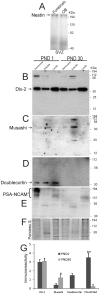
Figure 1. Western blots for neural stem cell and neuroprogenitor cell proteins in piglet forebrain.
A. Nestin immunoreactivity in forebrain subventricular zone (SVZ) and olfactory bulb (OB) SVZ. Lower four blots show immunoreactivities for Dlx2 (B), musashi (C), doublecortin (D), and PSA-NCAM (E) in OB SVZ subcellular extracts (membrane, soluble and crude nuclear) at postnatal day (PND) 1 and PND 30. Molecular weight standards (in kDa) are shown at right side of the blots. F. Representative Ponceau S-stained nitrocellulose membrane is shown for protein loading. G. Graph showing the densitometry quantification of the levels of immunoreactivity for Dlx-2, musashi, doublecortin, and PSa-NCAM in membrane (Dlx-2 and PSA-NCAM) and soluble (musashi and doublecortin) fractions in the OB periventricular region at PND1 and PND30. Values are mean ± SD. Single asterisk denotes musashi PND30 vs PND1 p < 0.05. Double asterisk denotes PSA-NCAM PND1 vs PND30 p < 0.001.
Figure 2
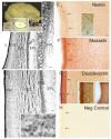
Figure 2. Piglet brain olfactory bulb (OB) gross anatomy, histology, and expression of neural stem cell and neuroprogenitor cell proteins.
A. Gross neuroanatomy of the 5-day-old piglet brain showing the gyrencephalic cortex, primary olfactory cortex (PC, also known as piriform cortex) and OB. Inset shows a Nissl-stained transverse section through the OB revealing the patent cerebroventricular cavity (V) and prominent subventricular zone (dark blue lining of the ventricle) containing the ependymal and subependymal layers. Scale bar = 1.1 mm. B. Nissl-stained transverse section revealing the architectural lamination of the OB. From the ventricle (V) outward the layers are: the SVZ, the granule cell layer (GCL), the mitral cell layer (MCL, the mitral cells project to the primary olfactory cortex), the external plexiform layer (EPL), the glomerular layer (GL), and the nerve fiber layer (NFL) that originates from the neuroepithelium of the olfactory mucosa. Scale bar = 100 µm. C. Higher magnification image showing the cellular detail of the wall of the OB. Scale bar = 33 µm. D. Nissl staining reveals the presence of mitotic figures (arrow) in the SVZ. Scale bar = 8.3 µm. E-G. Immunohistochemical localization of nestin (E), musashi (F), and doublecortin (G) in the OB. Immunoreactivity is seen as orange-brown staining. Arrows identify the subependymal layer. Open arrows (in E) identify nestin+ cells. Inset in E shows nestin+ cells (open arrows) at higher magnification. The large nucleus (pale oval) and sparse cytoplasm with prominent neurites is typical of a NSC. Insets in G show doublecortin immunoreactivity at the ependymal/subependymal layers (see box in G) at higher magnification in piglet OB at postnatal day (PND) 5 and 30. In PND5 inset the open arrow identifies doublecortin+ processes extending into the ependymal layer and hatched arrows identify doublecortin+ cell bodies in the subependymal layer. Scale bar in E (same for F-H) = 33 µm. Scale bars for insets = 12 µm (E) and 20 µm (G) H. Nestin primary antibody was preadsorbed against nestin synthetic peptide for a negative control. No staining is seen.
Figure 3
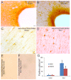
Figure 3. Localization of NSC and NPC markers in piglet forebrain SVZ and white matter.
A and B. Immunohistochemical localization of nestin (A) and musashi (B) shows enrichment of these two NSC markers in piglet forebrain SVZ. Immunoreactivity is seen as orange-brown staining. Arrows identify the SVZ. The lateral ventricle is seen at right. The opaque layer at the interface of the SVZ and the ventricular cavity is the ciliated ependymal layer. Scale bar = 33 µm. C and D. Immunohistochemical localization of nestin (C) and Dlx2 (D), a neuroprogenitor marker, shows enrichment in individual cells (arrows) in piglet subcortical white matter. Scale bar in C (same for D-F) = 17 µm. E and F. Nestin primary antibody (E) was preadsorbed against nestin synthetic peptide, and Dlx2 primary antibody (F) was preadsorbed against recombinant Dlx2. No staining is seen in either preparation. G. Graph showing the number of nestin+ and Dlx2+ cell bodies in the subcortical white matter of the parasagittal gyrus in primary somatosensory cortex in piglets at postnatal day (PND) 5 and 30. Values are mean ± SD (n= 3-4 piglets/group). Asterisk denotes significant difference (p < 0.05) compared to PND5.
Figure 4
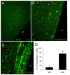
Figure 4. Bromodeoxyuridine (BrdU) cell proliferation tracking in piglet OB SVZ.
A and B. Confocal microscope images showing that the OB contains numerous BrdU+ cells (A, green) and subsets of these cells are newly born neurons as shown by the colocalization (B, seen as yellow, arrows) with the neuron marker NeuN. C. Confocal microscope images showing the co-labeling (arrows) of the DNA synthesis marker BrdU (red) and glial fibrillary acidic protein (GFAP, green) in the OB SVZ. Some BrdU+ nuclei are associated with GFAP+ cytoplasm. Scale bars = 33 µm (A), 17 µm (B), 20 µm (C). D. Graph showing the proportions of the total BrdU+ cells that are either GFAP+ or NeuN+. Values are mean ± SD (n=4). Some BrdU+ cells were not positive for either marker. Asterisk denotes NeuN significant difference (p < 0.001) compared to GFAP.
Figure 5
Figure 5. Bromodeoxyuridine (BrdU) cell proliferation tracking in piglet forebrain SVZ.
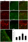
Confocal microscope images showing that subsets of BrdU+ cells in the forebrain SVZ can be immunophenotyped (arrows) as cells positive for the neuroblast marker doublecortin (A-C, Dcx), the astrocyte marker GFAP (D-F), the oligodendrocyte marker Olig2 (G-I), and the neuron marker NeuN (J). The lateral ventricle is identified (v). Scale bars: (in I, applies to A-I) = 12 µm; (in J) = 23 µm. K. Graph showing the proportions of the total BrdU+ cells that are either GFAP+ Olig2+, Dcx+, or NeuN+. Values are mean ± SD (n=4). Asterisk denotes Dcx significant difference (p < 0.05) compared to GFAP, Olig2, and NeuN.
Figure 6
Figure 6. In vivo BrdU labeling in piglet forebrain reveals a new corridor of newly born cells named the lateral migratory stream.
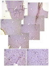
A-H. Images of a piglet forebrain section are shown with BrdU incorporation detected by the immunoperoxidase method (brown labeling) and counterstaining with cresyl violet. A low magnification image of piglet ventral forebrain is shown for perspective (A). Regions delineated by black rectangles in A are shown as higher magnification montage images in B-F that illustrate the distributions of BrdU labeled cells (hatched arrows, brown dots). Rostral migratory stream (RMS), and lateral ventricle (lv).G and H. Higher magnification images showing BrdU+ cell labeling (hatched arrows, brown nuclei) in the lateral migratory stream (G) beneath the striatum and in the piriform cortex (H). Unlabeled nuclei are pale violet. Scale bars: A = 300 µm, B (same for C-F) = 100 µm, G (same for H) = 25 µm.
Figure 7
Figure 7. Characterization of the lateral migratory stream (LMS) of newly born neurons and identification of immature neurons in piglet forebrain.
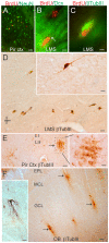
A. Confocal image showing the colocalization of BrdU (red) and NeuN (green) in subsets of neurons in the piriform cortex. Single-labeled neurons are green, single-labeled BrdU+ nuclei are red, and double-labeled neurons are yellow. B. Immunofluorescence showing the colocalization of BrdU (red) and doublecortin (Dcx, green) in the LMS. One Brdu+ cell (top cell) does not express Dcx, but another Brdu+ cell (bottom cell) is Dcx+ as seen by the yellow around the nucleus and the trailing Dcx-labeled process (arrow). C. Immunofluorescence showing the colocalization of BrdU (red) and β-tubulin III (βTubIII, green) in the LMS. D. Immmunoperoxidase staining for β-tubulin III (βTubIII) revealed a column of βTubIII+ immature neurons (brown cells) below the anterior striatum within the LMS. Inset shows a βTubIII+ LMS neuron with a fusiform morphology and leading and trailing processes. E. In piriform cortex, βTubIII+ immature neurons were found in cellular islands in layer II (arrow). Boxed area is shown at right at higher magnification and rotated 90°. F. In OB, βTubIII+ immature neurons (arrows) were found in the granule cell layer (GCL). These cells possessed extensive local vertical-oriented arborizations (inset) consistent with an interneuron morphology [46]. EPL, external plexiform layer; MCL, mitral cell layer. Scale bars = 25 µm (A), 5 µm (B), 4 µm (C), 25 µm (D), 8 µm (D inset), 32 µm (E), 12.5 µm (E right), 100 µm (F), 25 µm (F inset).
Figure 8
Figure 8. Newborn piglet OB-SVZ cells form multipotent neurospheres.
A. Floating neurosphere formed by notch-1-immunopanned NSCs isolated from the OB. Scale = 12 µm. B-D. Attached neurospheres dispersed as monolayers are multipotent by differentiating into cells that are neurons identified by MAP2, astrocytes identified by GFAP, and oligodendrocytes identified by O4. E. Neurosphere density increased when medium was supplemented with FGF2. Scale bars = 12.5 (B), 20 (C), 20 (D), 50 (E) µm.
Figure 9
Figure 9. Single-cell clonal analysis of piglet OB- and forebrain-SVZ NSCs and genetic tagging with green fluorescent protein (GFP).
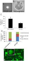
A. A single cell isolated from a neurosphere derived from the OB SVZ. Scale bar = 12 µm. B. Secondary neurosphere formed from neurosphere-derived single cells. Scale bar = 12 µm. C and D. FGF2/EGF-stimulated growth assays on dissociated neurosphere cells isolated from the forebrain-SVZ and OB-SVZ. The number of secondary neurospheres formed (C, values are mean ± SD) and individual neurosphere cell number (D) were assessed. Asterisk denotes SVZ-forebrain significant difference (p < 0.01) compared to SVZ-OB. E. Numerous piglet OB-SVZ-derived neurospheres were robustly and stably transduced to express GFP by lentiviral gene transfer. Inset shows a single GFP-tagged neurosphere at higher magnification. Some individual cells can be seen in the sphere. Scale bars: 80 µm, 5 µm (inset).

Similar articles

Cited by

References

    1. Santner-Nanan B, Peek MJ, McCullagh P, Nanan R (2005) Therapeutic potential of stem cells in perinatal medicine. Aust N Z J Obstet Gynaecol 45: 102-107. PubMed: 15760308. - PubMed
    1. Titomanlio L, Kavelaars A, Dalous J, Mani S, El Ghouzzi V et al. (2011) Stem cell therapy for neonatal brain injury: perspectives and challenges. Ann Neurol 70: 698-712. doi:10.1002/ana.22518. PubMed: 22162055. - DOI - PubMed
    1. Ikeda T, Iwai M, Hayashi T, Nagano I, Shogi M et al. (2005) Limited differentiation to neurons and astroglia from neural stem cells in the cortex and striatum after ischemia/hypoxia in the neonatal rat brain. Am J Obstet Gynecol 193: 849-856. doi:10.1016/j.ajog.2005.01.029. PubMed: 16150285. - DOI - PubMed
    1. Park KI, Teng YD, Snyder EY (2002) The injured brain interacts reciprocally with neural stem cells supported by scaffolds to reconstitute lost tissue. Nat Biotechnol 20: 1111-1117. doi:10.1038/nbt751. PubMed: 12379868. - DOI - PubMed
    1. Zheng T, Rossignol C, Leibovici A, Anderson KJ, Steindler DA et al. (2006) Transplantation of multipotent astrocytic stem cells into a rat model of neonatal hypoxic-ischemic encephalopathy. Brain Res 1112: 99-105. doi:10.1016/j.brainres.2006.07.014. PubMed: 16919606. - DOI - PubMed

Publication types

LinkOut - more resources