

MicroSPECT and MicroPET Imaging of Small Animals for Drug Development
- PMID: 24278622
- PMCID: PMC3834443
- DOI: 10.5487/TR.2013.29.1.001
MicroSPECT and MicroPET Imaging of Small Animals for Drug Development
Abstract
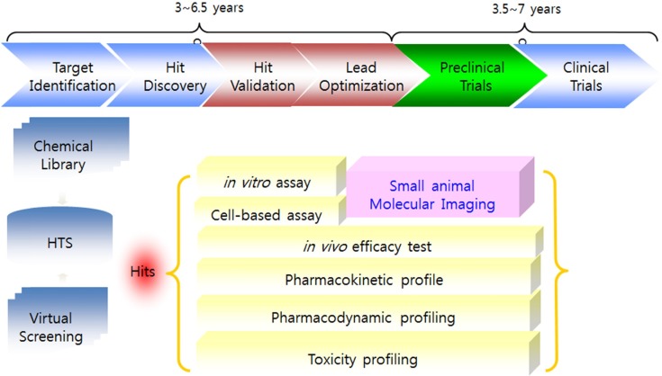
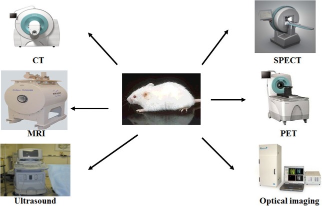
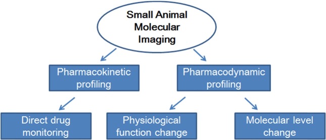
The process of drug discovery and development requires substantial resources and time. The drug industry has tried to reduce costs by conducting appropriate animal studies together with molecular biological and genetic analyses. Basic science research has been limited to in vitro studies of cellular processes and ex vivo tissue examination using suitable animal models of disease. However, in the past two decades new technologies have been developed that permit the imaging of live animals using radiotracer emission, Xrays, magnetic resonance signals, fluorescence, and bioluminescence. The main objective of this review is to provide an overview of small animal molecular imaging, with a focus on nuclear imaging (single photon emission computed tomography and positron emission tomography). These technologies permit visualization of toxicodynamics as well as toxicity to specific organs by directly monitoring drug accumulation and assessing physiological and/or molecular alterations. Nuclear imaging technology has great potential for improving the efficiency of the drug development process.
Keywords: Animal experimental model; Drug development; Molecular imaging; Nuclear imaging; microPET; microSPECT.
Figures
References
-
- DiMasi J.A., Hansen R.W., Grabowski H.G. The price of innovation: new estimates of drug development costs. J. Health Econ. (2003);22:151–185. - PubMed
-
- Ahn B.C. Applications of molecular imaging in drug discovery and development process. Curr. Opin. Biotechnol. (2011);12:459–468. - PubMed
-
- Rudin M. Noninvasive structural, fuctional, and molecular imaging in drug development. Curr. Opin. Chem. Biol. (2009);13:360–371. - PubMed
-
- Czernin J., Weber W.A., Herschman H.R. Molecular imaging in development of cancer therapeutics. Annu. Rev. Med. (2006);57:99–118. - PubMed
-
- Rudin M., Weissleder R. Molecular imaging in drug discovery and development. Nat. Rev. Drug Discov. (2003);2:123–131. - PubMed
Publication types
LinkOut - more resources
Full Text Sources
Other Literature Sources
Miscellaneous
