

Malignant solitary fibrous tumor of tandem lesions in the skull and spine
- PMID: 24278657
- PMCID: PMC3836935
- DOI: 10.3340/jkns.2013.54.3.246
Malignant solitary fibrous tumor of tandem lesions in the skull and spine
Abstract
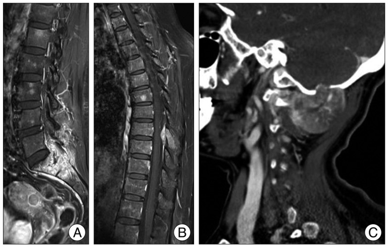
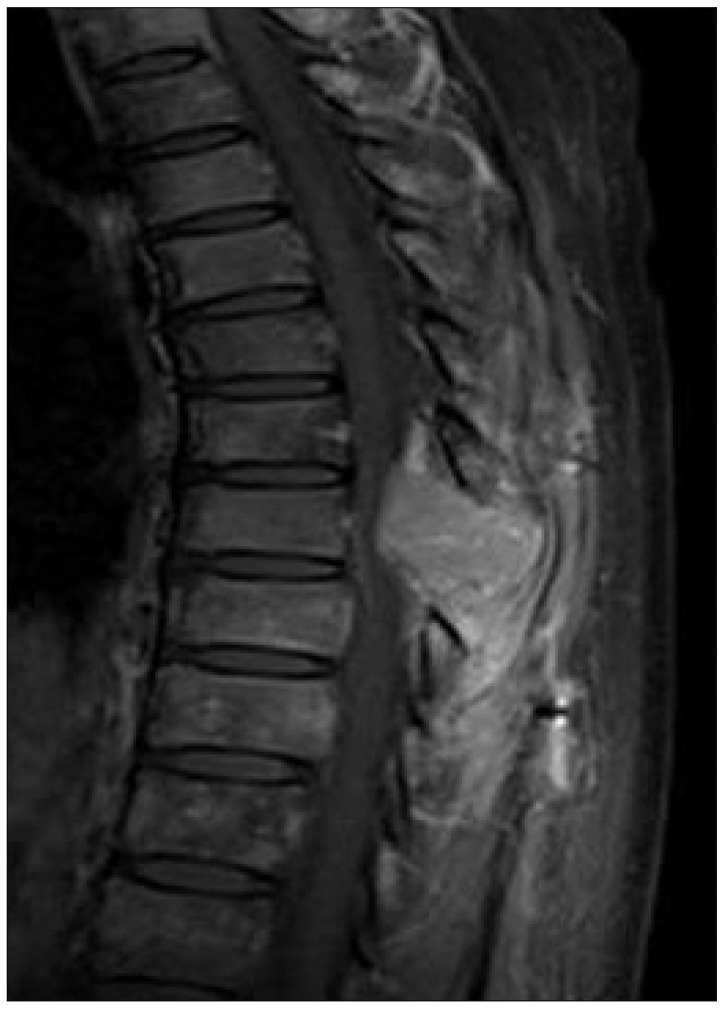
A Solitary Fibrous Tumor (Sft) Is A Rare Neoplasm Originated From The Pleura, But They Can Occur In A Variety Of Extrathoracic Regions. Although Many Cases Of Primary Sft Have Been Reported, There Are Extremely Rare Repots To Date Of A Malignant Sft In The Spine Or Skull. A 54-year-woman Visited Our Hospital Due To Low Back Pain And Both Leg Radiating Pain. Several Imaging Studies Including Magnetic Resonance Imaging And Computed Tomography Revealed Expansive Enhanced Lesions In The Occipital Bone, T8, S1-2, And Ilium, With Neural Tissue Compression. We Performed Surgical Resection Of The Tumor In Each Site, And Postoperative Radiosurgery And Chemotherapy Were Performed. However, After Six Months, Tumors Were Recurred And Metastasized In Multiple Regions Including Whole Spine And Lung. The Authors Report Here The First Case Of Patient With Malignant Sft Of Tandem Lesions In The Various Bony Structures, Including Skull, Thoracic Spine, And Sacral Spine, With A Rapid Recurrence And Metastasis. Although Malignant Sft Is Extremely Rare, It Should Be Considered In The Differential Diagnosis And Carful Follow-up Is Needed.
Keywords: Metastasis; Skull; Solitary fibrous tumors; Spine.
Figures

References
-
- Briselli M, Mark EJ, Dickersin GR. Solitary fibrous tumors of the pleura : eight new cases and review of 360 cases in the literature. Cancer. 1981;47:2678–2689. - PubMed
-
- Chan JK. Solitary fibrous tumour--everywhere, and a diagnosis in vogue. Histopathology. 1997;31:568–576. - PubMed
-
- Cox DP, Daniels T, Jordan RC. Solitary fibrous tumor of the head and neck. Oral Surg Oral Med Oral Pathol Oral Radiol Endod. 2010;110:79–84. - PubMed
-
- Demicco EG, Park MS, Araujo DM, Fox PS, Bassett RL, Pollock RE, et al. Solitary fibrous tumor : a clinicopathological study of 110 cases and proposed risk assessment model. Mod Pathol. 2012;25:1298–1306. - PubMed
Publication types
LinkOut - more resources
Full Text Sources
Other Literature Sources
