

Spinal Hemangiopericytoma Which Needed Intraoperative Embolization due to Unexpected Bleeding
- PMID: 24278659
- PMCID: PMC3836937
- DOI: 10.3340/jkns.2013.54.3.253
Spinal Hemangiopericytoma Which Needed Intraoperative Embolization due to Unexpected Bleeding
Abstract
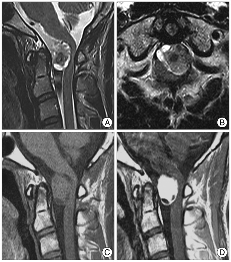
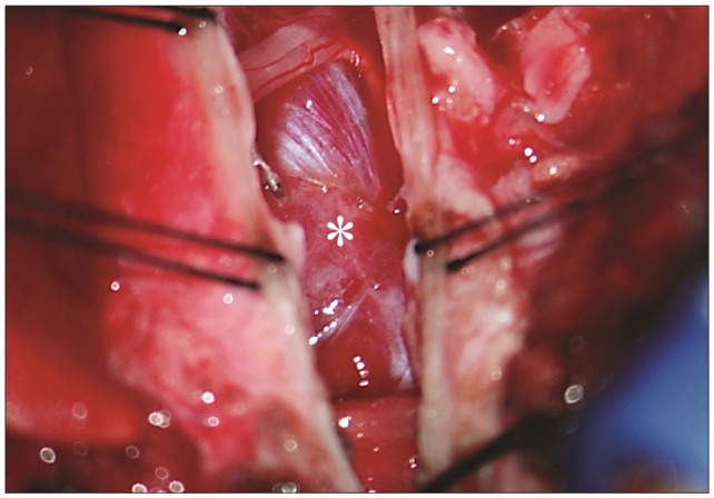
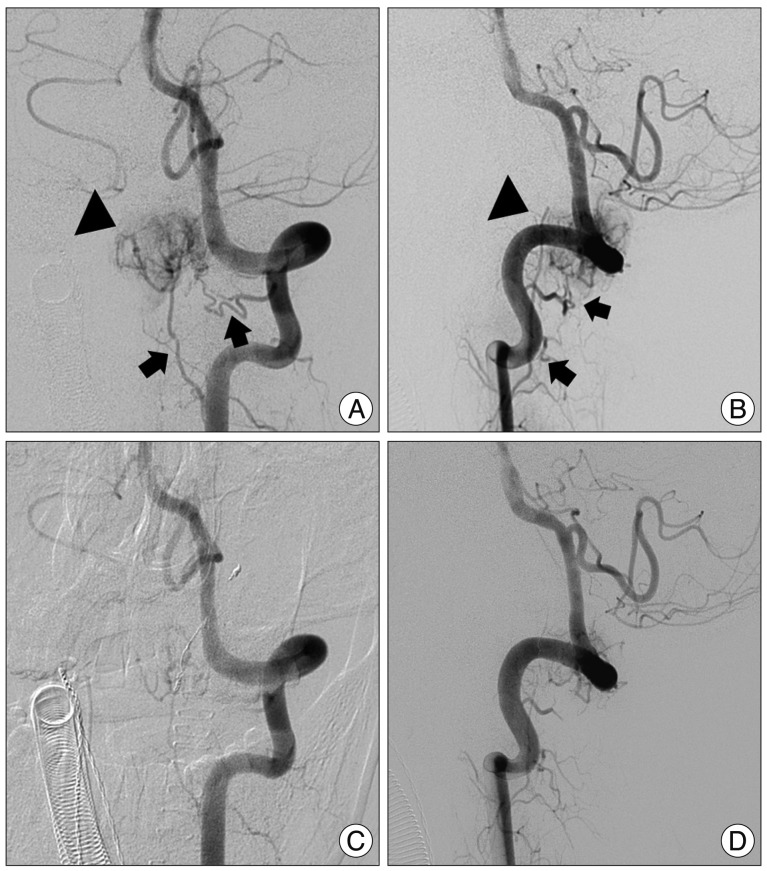
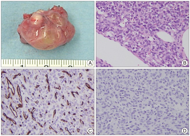
Spinal intradural hemangiopericytoma is a very rare tumor and can be characterized by massive bleeding during surgeries, frequent recurrence, and metastasis. However, definite radiologic differential points of hemangiopericytoma are not known. We describe an unexpected hemangiopericytoma case with large bleeding and management of the tumor. A 21-year-old man visited complaining of progressive neck pain and tingling sensation in both hands. Magnetic resonance imaging of his spine revealed C1-2 ventral intradural mass. When the dura was opened, the intradural tumor was placed behind spinal accessary nerves. The tumor was partially exposed only after some accessary nerves had been cut. When internal debulking was performing, unexpected bleeding was noted and it was difficult to control because of narrow surgical field and hypervascularity. Intraoperative spinal angiography and embolization were performed. The tumor was completely removed after embolization. Pathological diagnosis was consistent with hemangiopericytoma. When surgeons meet a flesh-red tumor that bleeds unexpectedly during surgery, hemangiopericytoma may be considered. When feeder control is hard due to reciprocal location of spinal cord, the tumor, and feeders, intraoperative angiography and embolization may be a possible option.
Keywords: Angiography; Hemangiopericytoma; Intradural; Spine; Surgery.
Figures

References
-
- Ackerman PD, Khaldi A, Shea JF. Intradural hemangiopericytoma of the thoracic spine : a case report. Spine J. 2011;11:e9–e14. - PubMed
-
- Betchen S, Schwartz A, Black C, Post K. Intradural hemangiopericytoma of the lumbar spine : case report. Neurosurgery. 2002;50:654–657. - PubMed
-
- Ciappetta P, Celli P, Palma L, Mariottini A. Intraspinal hemangiopericytomas Report of two cases and review of the literature. Spine (Phila Pa 1976) 1985;10:27–31. - PubMed
-
- Dufour H, Métellus P, Fuentes S, Murracciole X, Régis J, Figarella-Branger D, et al. Meningeal hemangiopericytoma : a retrospective study of 21 patients with special review of postoperative external radiotherapy. Neurosurgery. 2001;48:756–762. discussion 762-763. - PubMed
Publication types
LinkOut - more resources
Full Text Sources
Other Literature Sources
