

Genetic aspects of congenital and idiopathic scoliosis
- PMID: 24278672
- PMCID: PMC3820596
- DOI: 10.6064/2012/152365
Genetic aspects of congenital and idiopathic scoliosis
Abstract
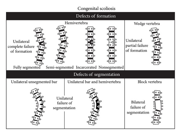
Congenital and idiopathic scoliosis represent disabling conditions of the spine. While congenital scoliosis (CS) is caused by morphogenic abnormalities in vertebral development, the cause(s) for idiopathic scoliosis is (are) likely to be varied, representing alterations in skeletal growth, neuromuscular imbalances, disturbances involving communication between the brain and spine, and others. Both conditions are characterized by phenotypic and genetic heterogeneities, which contribute to the difficulties in understanding their genetic basis that investigators face. Despite the differences between these two conditions there is observational and experimental evidence supporting common genetic mechanisms. This paper focuses on the clinical features of both CS and IS and highlights genetic and environmental factors which contribute to their occurrence. It is anticipated that emerging genetic technologies and improvements in phenotypic stratification of both conditions will facilitate improved understanding of the genetic basis for these conditions and enable targeted prevention and treatment strategies.
Figures
References
-
- Riseborough EJ, Wynne Davies R. A genetic survey of idiopathic scoliosis in Boston, Massachusetts. Journal of Bone and Joint Surgery—Series A. 1973;55(5):974–982. - PubMed
-
- Shands AR, Eisberg HB. The incidence of scoliosis in the state of Delaware; a study of 50,000 minifilms of the chest made during a survey for tuberculosis. Journal of Bone and Joint Surgery— Series A. 1955;37(6):1243–1249. - PubMed
-
- Rogala EJ, Drummond DS, Gurr J. Scoliosis: incidence and natural history. A prospective epidemiological study. Journal of Bone and Joint Surgery—Series A. 1978;60(2):173–176. - PubMed
-
- Brand MC. Examination of the newborn with congenital scoliosis: focus on the physical. Advances in Neonatal Care. 2008;8(5):265–273. - PubMed
-
- Davies BR, Durán M. Malformations of the cranium, vertebral column, and related central nervous system: morphologic heterogeneity may indicate biological diversity. Birth Defects Research Part A—Clinical and Molecular Teratology. 2003;67(8):563–571. - PubMed
Publication types
LinkOut - more resources
Full Text Sources
Medical
