Review
. 2012:2012:424965.
doi: 10.6064/2012/424965. Epub 2012 Aug 21.

The visual effects of intraocular colored filters

Affiliations
Review

The visual effects of intraocular colored filters

Billy R Hammond Jr. Scientifica (Cairo). 2012.

Abstract

Modern life is associated with a myriad of visual problems, most notably refractive conditions such as myopia. Human ingenuity has addressed such problems using strategies such as spectacle lenses or surgical correction. There are other visual problems, however, that have been present throughout our evolutionary history and are not as easily solved by simply correcting refractive error. These problems include issues like glare disability and discomfort arising from intraocular scatter, photostress with the associated transient loss in vision that arises from short intense light exposures, or the ability to see objects in the distance through a veil of atmospheric haze. One likely biological solution to these more long-standing problems has been the use of colored intraocular filters. Many species, especially diurnal, incorporate chromophores from numerous sources (e.g., often plant pigments called carotenoids) into ocular tissues to improve visual performance outdoors. This review summarizes information on the utility of such filters focusing on chromatic filtering by humans.


Figures

Figure 1
Figure 1
The photopic (light-adapted) spectral sensitivity function plotted next to the internal colored filters, the yellow crystalline lens, and macular pigment (from Wyszecki and Stiles, [64]). Note that the colored filters do not significantly overlap the photopic spectral sensitivity function.
Figure 2
Figure 2
The scotopic sensitivity curve plotted with MP and lens absorbance (Wyszecki and Stiles, [64]). This curve is significantly shifted to the short-wave end as compared to the photopic curve above. Note that the lens is still not a major filtering impediment to dark-adapted sensitivity (fortuitous since it screens rods). MP density, however, does significantly overlap with the curve. MP is localized within the retina as to mostly screen the cones and not the rods to obviate this problem.
Figure 3
Figure 3
The example above shows an achromatic test target and surround of nearly the same luminance. The central target becomes visible when either the target is just slightly brighter or the surround is just slightly darker. This very small change in brightness is enough to create a luminance edge (a phenomenon called brightness induction). These experiments are typically done with achromatic stimuli like those shown above but a similar effect would hold for colored stimuli.
Figure 4
Figure 4
This example shows a blue surround with a yellow stimulus of equal luminance. Note that what defines the edge is based only on the wavelength or color difference. The luminance difference itself, however, will be exaggerated by differential absorption by macular pigment.
Figure 5
Figure 5
Data for the rod and cone densities were obtained from the original data by Osterberg, 1935. The MP distribution was obtained from Werner et al. 2000 who measured MP density with HFP using a 12-degree reference. Note that for this example, MP is screening a significant number of rods in the central macula (around 10 degrees in diameter).
Figure 6
Figure 6
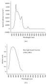
Light emitted by a computer monitor set to a blue background (a) compared to the blue-light hazard function published by ANSI.

References

    1. Tanito M, Okuno T, Ishiba Y, Ohira A. Transmission spectrums and retinal blue-light irradiance values of untinted and yellow-tinted intraocular lenses. Journal of Cataract and Refractive Surgery. 2010;36(2):299–307. - PubMed
    1. Mukai K, Matsushima H, Sawano M, Nobori H, Obara Y. Photoprotective effect of yellow-tinted intraocular lenses. Japanese Journal of Ophthalmology. 2009;53(1):47–51. - PubMed
    1. Hammond BR, Jr., Bernstein B, Dong J. The effect of the acrysof natural lens on glare disability and photostress. American Journal of Ophthalmology. 2009;148(2):272–e2. - PubMed
    1. Hammond BR, Jr., Renzi LM, Sachak S, Brint SF. Contralateral comparison of blue-filtering and non-blue-filtering intraocular lenses: glare disability, heterochromatic contrast, and photostress recovery. Clinical Ophthalmology. 2010;4(1):1465–1473. - PMC - PubMed
    1. Provines WF, Rahe AJ, Block MG, Pena T, Tredici TJ. Yellow lens effects upon visual acquisition performance. Aviation Space and Environmental Medicine. 1992;63(7):561–564. - PubMed

LinkOut - more resources