

Periventricular Heterotopia: Shuttling of Proteins through Vesicles and Actin in Cortical Development and Disease
- PMID: 24278701
- PMCID: PMC3820590
- DOI: 10.6064/2012/480129
Periventricular Heterotopia: Shuttling of Proteins through Vesicles and Actin in Cortical Development and Disease
Abstract
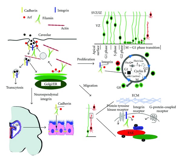
During cortical development, proliferating neural progenitors exhibit polarized apical and basolateral membranes that are maintained by tightly controlled and membrane-specific vesicular trafficking pathways. Disruption of polarity through impaired delivery of proteins can alter cell fate decisions and consequent expansion of the progenitor pool, as well as impact the integrity of the neuroependymal lining. Loss of neuroependymal integrity disrupts radial glial scaffolding and alters initial neuronal migration from the ventricular zone. Vesicle trafficking is also required for maintenance of lipid and protein cycling within the leading and trailing edge of migratory neurons, as well as dendrites and synapses of mature neurons. Defects in this transport machinery disrupt neuronal identity, migration, and connectivity and give rise to a malformation of cortical development termed as periventricular heterotopia (PH). PH is characterized by a reduction in brain size, ectopic clusters of neurons localized along the lateral ventricle, and epilepsy and dyslexia. These anatomical anomalies correlate with developmental impairments in neural progenitor proliferation and specification, migration from loss of neuroependymal integrity and neuronal motility, and aberrant neuronal process extension. Genes causal for PH regulate vesicle-mediated endocytosis along an actin cytoskeletal network. This paper explores the role of these dynamic processes in cortical development and disease.
Figures
References
-
- Bystron I, Blakemore C, Rakic P. Development of the human cerebral cortex: boulder Committee revisited. Nature Reviews Neuroscience. 2008;9(2):110–122. - PubMed
-
- Gotz M, Huttner WB. The cell biology of neurogenesis. Nature Reviews Molecular Cell Biology. 2005;6(10):777–788. - PubMed
-
- Rakic P. Specification of cerebral cortical areas. Science. 1988;241(4862):170–176. - PubMed
-
- Couillard-Despres S, Winkler J, Uyanik G, Aigner L. Molecular mechanisms of neuronal migration disorders, quo vadis? Current Molecular Medicine. 2001;1(6):677–688. - PubMed
-
- Huttner WB, Brand M. Asymmetric division and polarity of neuroepithelial cells. Current Opinion in Neurobiology. 1997;7(1):29–39. - PubMed
Publication types
LinkOut - more resources
Full Text Sources
Medical
