Review
. 2012:2012:703675.
doi: 10.6064/2012/703675. Epub 2012 Dec 19.

The neuropathology of autism

Affiliations
Review

The neuropathology of autism

Gene J Blatt. Scientifica (Cairo). 2012.

Abstract

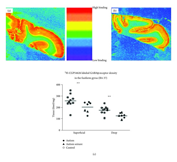
Autism is a behaviorally defined neurodevelopmental disorder that affects over 1% of new births in the United States and about 2% of boys. The etiologies are unknown and they are genetically complex. There may be epigenetic effects, environmental influences, and other factors that contribute to the mechanisms and affected neural pathway(s). The underlying neuropathology of the disorder has been evolving in the literature to include specific brain areas in the cerebellum, limbic system, and cortex. Part(s) of structures appear to be affected most rather than the entire structure, for example, select nuclei of the amygdala, the fusiform face area, and so forth. Altered cortical organization characterized by more frequent and narrower minicolumns and early overgrowth of the frontal portion of the brain, affects connectivity. Abnormalities include cytoarchitectonic laminar differences, excess white matter neurons, decreased numbers of GABAergic cerebellar Purkinje cells, and other events that can be traced developmentally and cause anomalies in circuitry. Problems with neurotransmission are evident by recent receptor and binding site studies especially in the inhibitory GABA system likely contributing to an imbalance of excitatory/inhibitory transmission. As postmortem findings are related to core behavior symptoms, and technology improves, researchers are gaining a much better perspective of contributing factors to the disorder.


Figures

Figure 1
Figure 1
Scatter plot for neuronal number in the inferior olivary complex by group. No significant differences were found for the IOC (DAO: dorsal accessory olive; MAO: medial accessory olive; PO: principle olive). A: autism, C: control.
Figure 2
Figure 2
Photomicrographs of the inferior olive to show clustering at the edge of principle olive in an autistic brain. In A (autism) the clustering of neurons can be seen as a single row at the edge of the principle olive (arrow), whereas in B (control) these neurons are more scattered although some neurons do exist on the edge of the ribbon (arrow). Scale bars = 200 μm.
Figure 3
Figure 3
Scatter plot showing the perikaryal volume for the inferior olivary complex by group (DAO: dorsal accessory olive; MAO: medial accessory olive; PO: principle olive). Although there were no significant differences found, the PO neuronal volume was quite variable in both autistic and control brains. A: autism, C: control.
Figure 4
Figure 4
Photomicrographs showing similar altered formations of the principle olive in the autistic (A and C) and control brains (B and D). A and B show what appear to be reduplications (arrows) and C and D show examples of a gap in the ribbon (arrows) of the principle olive which may be caused by perforating vessel(s).
Figure 5
Figure 5
GAD65 mRNA-labeled neurons (arrows) in the dentate nuclei in a control case illustrating a small cell in A and a larger cell in B. Note the silver grain GAD65-mRNA-positive labeling throughout both cells. Scale bar in A = 20 μm and refers to both A and B. Permissions granted from modified figure found in Yip et al., 2009 [47].
Figure 6
Figure 6
Lamination of ACC in control and autism brains (40x). Note irregular lamination obvious in layers III-V in an autism case (left figure) compared to control at right. In each figure, layer II is on right side, layers V-Vi at left. Also note, the ACC lacks a layer IV. Permissions granted for modified figure from Simms et al., 2009 [81]. Scale bar = 1 mm.
Figure 7
Figure 7
A 50 μm vibratome cut section demonstrates vertical minicolumnar organization in the human inferior frontal cortex via immunohistochemistry section incubated with primary antibodies to microtubule-associated protein type 2 (MAP2) that labels apical dendrites (Figure 1; 1000x; red circle). Note the radial organization of columnar components in this tangential section through layer IV. This represents the center of a cortical minicolumn.
Figure 8
Figure 8
Graph demonstrating [3H]-CGP54626 labeled GABAB receptor binding density in the fusiform gyrus (FG) from a control (a) and an autistic case (b). (c) is a scatter plot of all cases included in the study. Significant reductions (∗∗) in the superficial (P = 0.019) and deep (P = 0.00095) layers were found in the autism cases compared to age- and postmortem interval-matched controls. Permissions granted from Oblak et al., 2010 [95].

References

    1. Kanner L. Autistic disturbances of affective contact. The Nervous Child. 1943;2:217–250.
    1. Bauman M, Kemper TL. Histoanatomic observations of the brain in early infantile autism. Neurology. 1985;35(6):866–874. - PubMed
    1. Bauman ML, Kemper TL. Neuroanatomic observations of the brain in autism. In: Bauman ML, Kemper TL, editors. The Neurobiology of Autism. Baltimore, Md, USA: Johns Hopkins University Press; 1994. pp. 119–145.
    1. Bailey A, Phillips W, Rutter M. Autism: towards an integration of clinical, genetic, neuropsychological, and neurobiological perspectives. Journal of Child Psychology and Psychiatry and Allied Disciplines. 1996;37(1):89–126. - PubMed
    1. Bailey A, Luthert P, Dean A, et al. A clinicopathological study of autism. Brain. 1998;121(5):889–905. - PubMed