Review
. 2013:2013:675213.
doi: 10.1155/2013/675213. Epub 2013 Jan 9.

Global Molecular Epidemiology of Cryptococcus neoformans and Cryptococcus gattii: An Atlas of the Molecular Types

Affiliations
Review

Global Molecular Epidemiology of Cryptococcus neoformans and Cryptococcus gattii: An Atlas of the Molecular Types

Massimo Cogliati. Scientifica (Cairo). 2013.

Abstract

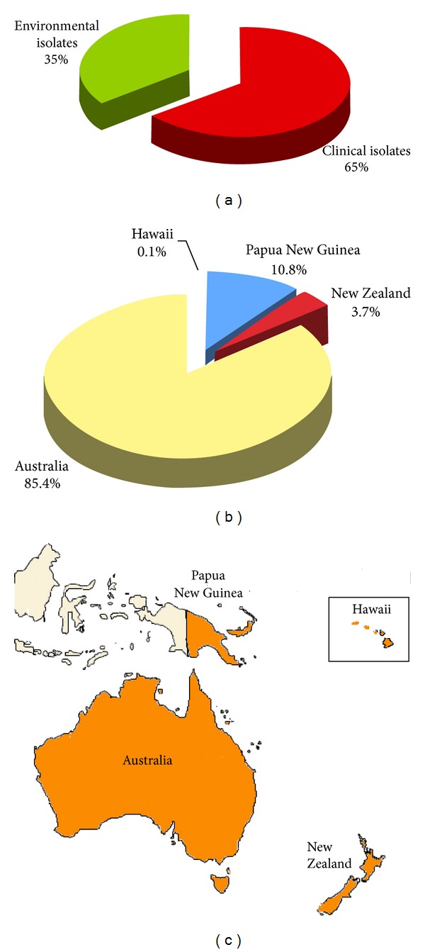
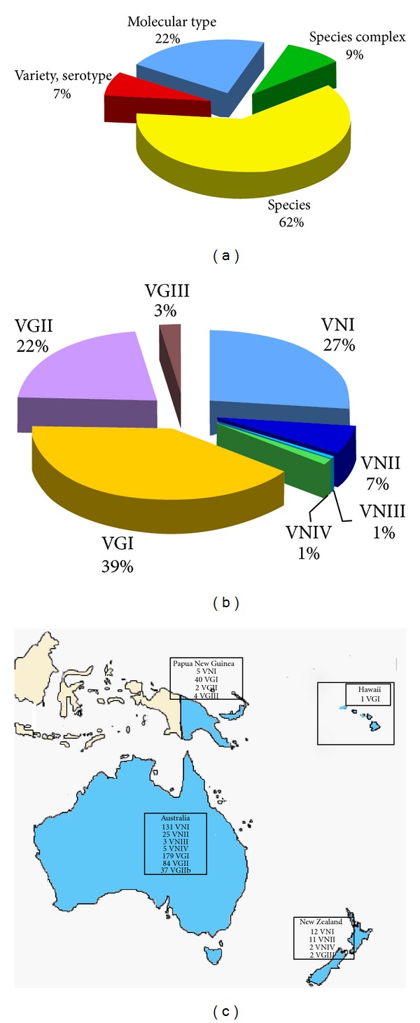
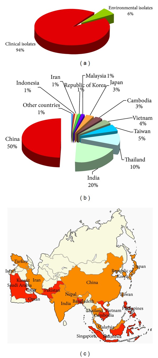
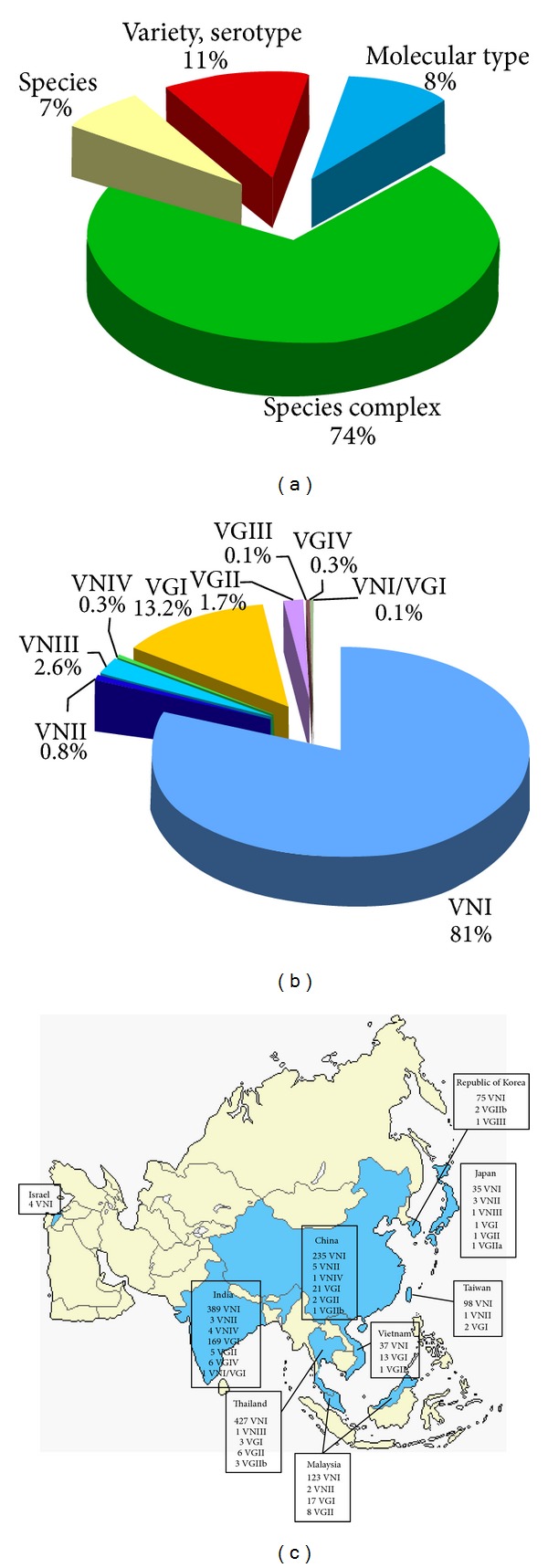
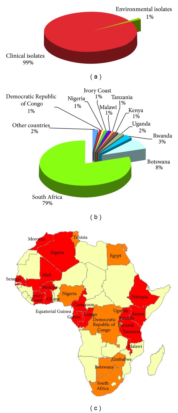
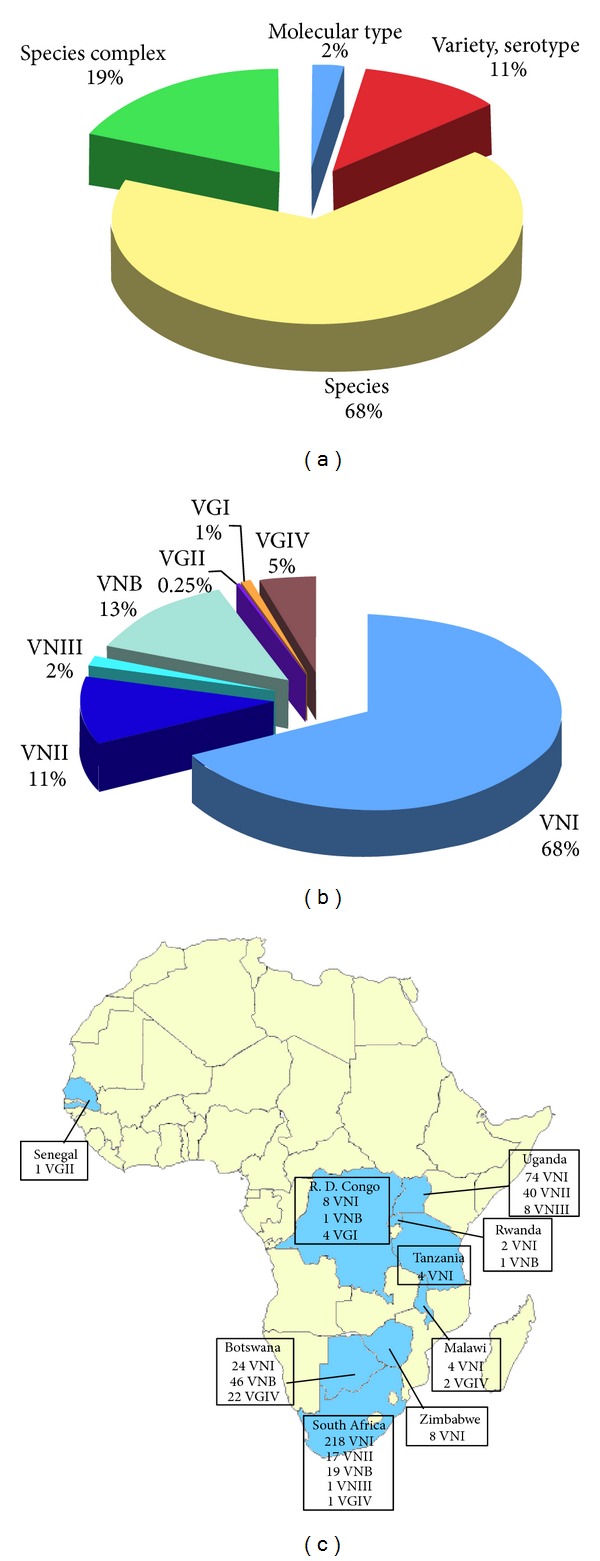
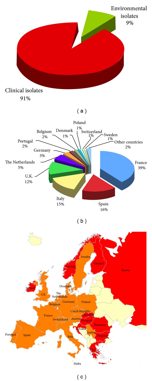
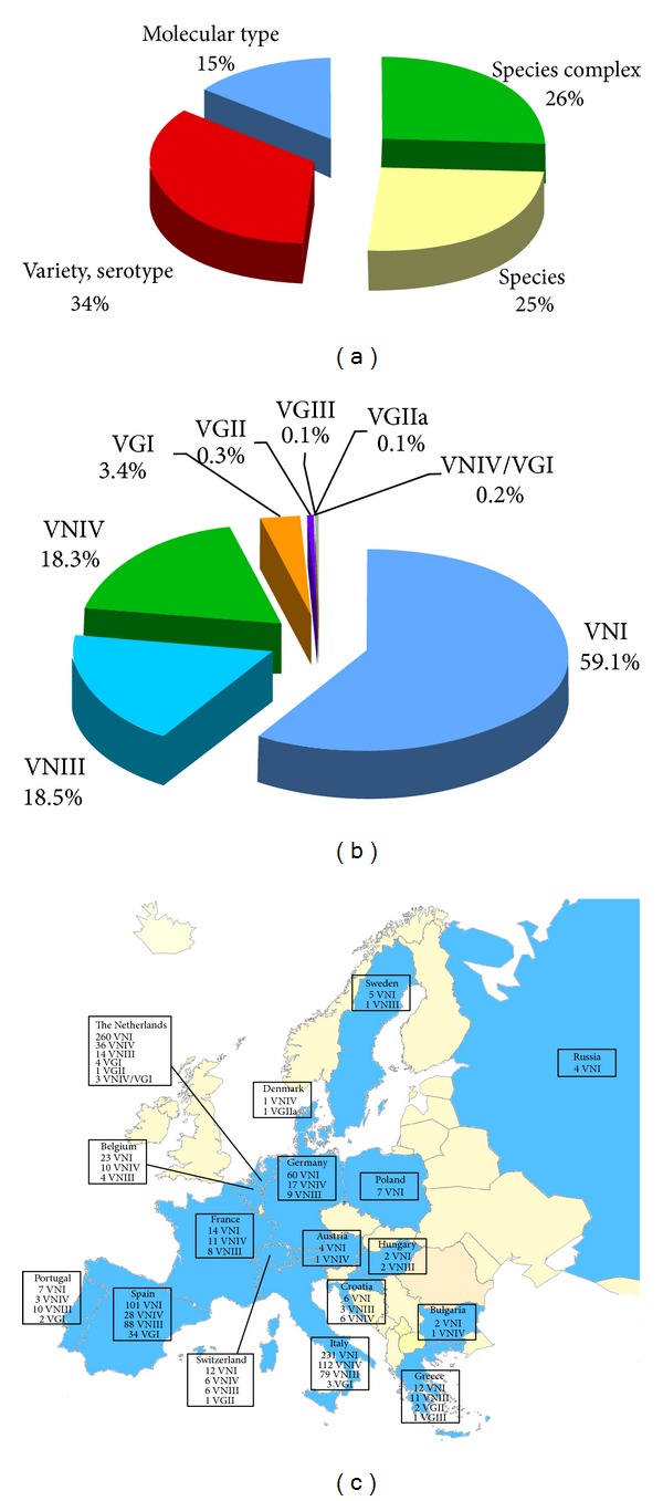
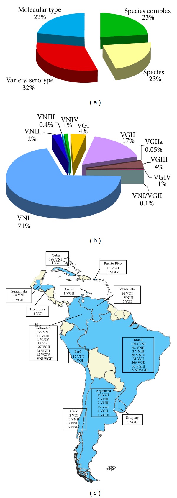
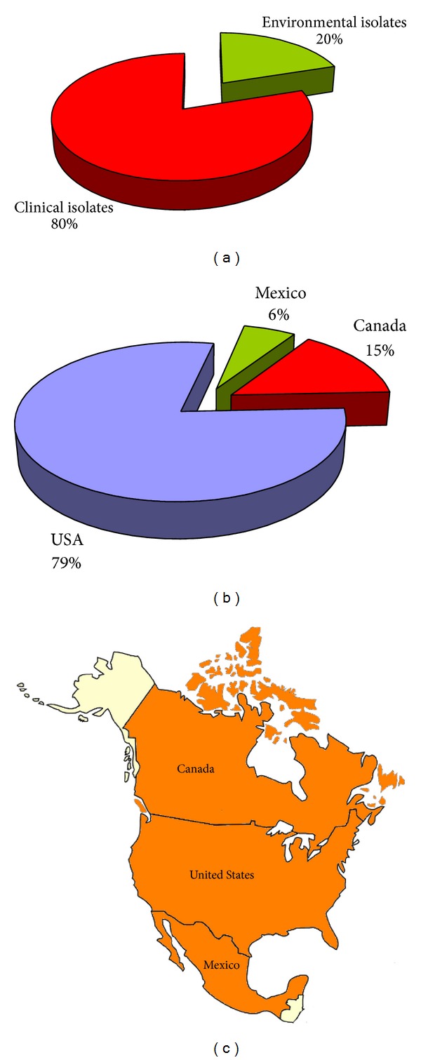
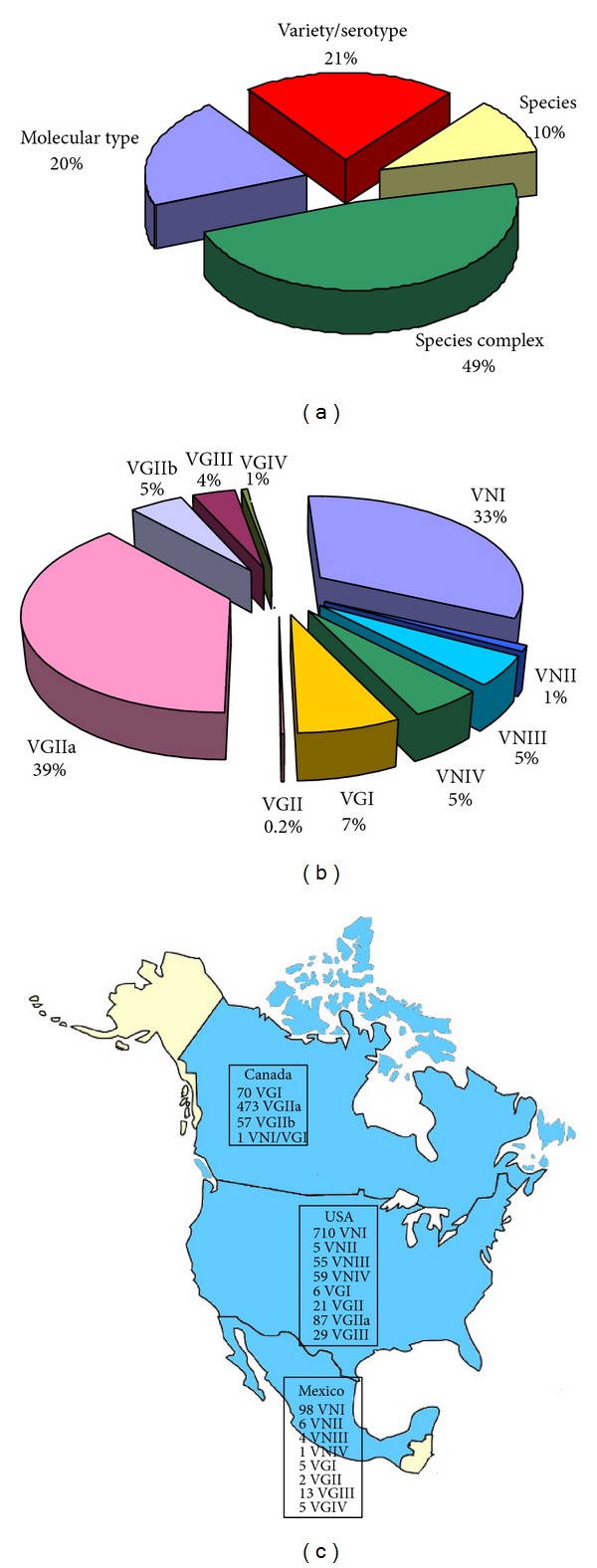
Cryptococcosis is a fungal disease affecting more than one million people per year worldwide. The main etiological agents of cryptococcosis are the two sibling species Cryptococcus neoformans and Cryptococcus gattii that present numerous differences in geographical distribution, ecological niches, epidemiology, pathobiology, clinical presentation and molecular characters. Genotyping of the two Cryptococcus species at subspecies level supplies relevant information to understand how this fungus has spread worldwide, the nature of its population structure, and how it evolved to be a deadly pathogen. At present, nine major molecular types have been recognized: VNI, VNII, VNB, VNIII, and VNIV among C. neoformans isolates, and VGI, VGII, VGIII, and VGIV among C. gattii isolates. In this paper all the information available in the literature concerning the isolation of the two Cryptococcus species has been collected and analyzed on the basis of their geographical origin, source of isolation, level of identification, species, and molecular type. A detailed analysis of the geographical distribution of the major molecular types in each continent has been described and represented on thematic maps. This study represents a useful tool to start new epidemiological surveys on the basis of the present knowledge.


Figures

Figure 1
Figure 1
Percentage of Cryptococcus neoformans and Cryptococcus gattii isolates from clinical (n = 1,594) and environmental (n = 924) sources (a). Distribution of Cryptococcus neoformans and Cryptococcus gattii isolates in the different countries of Oceania (b). Map of the geographical distribution of the Cryptococcus neoformans and Cryptococcus gattii isolates in Oceania (c). Clinical isolates were reported from red-colored countries, whereas both clinical and environmental isolates were reported from orange-colored countries.
Figure 2
Figure 2
Percentage of Cryptococcus neoformans and Cryptococcus gattii isolates (n = 2,518) identified at species complex, species, variety/serotype, or molecular type level (a). Prevalence of the different VN and VG molecular types among the isolates identified at molecular type level (n = 543) (b). Geographic distribution of the molecular types identified in Oceania (c). Molecular typing data have been combined from the following references: Australia [–27], New Zealand [13, 15], and Papua New Guinea [–15, 23, 25].
Figure 3
Figure 3
Percentage of Cryptococcus neoformans and Cryptococcus gattii isolates from clinical (n = 18,412) and environmental (n = 1,239) sources (a). Distribution of Cryptococcus neoformans and Cryptococcus gattii isolates in the different Asian countries (b). Map of the geographical distribution of the Cryptococcus neoformans and Cryptococcus gattii isolates in Asia (c). Clinical isolates were reported from red-colored countries, whereas both clinical and environmental isolates were reported from orange-colored countries.
Figure 4
Figure 4
Percentage of Cryptococcus neoformans and Cryptococcus gattii isolates (n = 19,651) identified at species complex, species, variety/serotype, or molecular type level (a). Prevalence of the different VN and VG molecular types among the isolates identified at molecular type level (n = 1,708) (b). Geographic distribution of the molecular types identified in Asia (c). Molecular typing data have been combined from the following references: India [, , , –36], China [, –43], Thailand [, , –48], Malaysia [49, 50], Vietnam [51], Taiwan [38, 52], Japan [38, 53, 54], Republic of Korea [55], and Israel [56].
Figure 5
Figure 5
Percentage of Cryptococcus neoformans and Cryptococcus gattii isolates from clinical (n = 19,436) and environmental (n = 211) sources (a). Distribution of Cryptococcus neoformans and Cryptococcus gattii isolates in the different African countries (b). Map of the geographical distribution of the Cryptococcus neoformans and Cryptococcus gattii isolates in Africa (c). Clinical isolates were reported from red-colored countries, whereas both clinical and environmental isolates were reported from orange-colored countries.
Figure 6
Figure 6
Percentage of Cryptococcus neoformans and Cryptococcus gattii isolates (n = 19,647) identified at species complex, species, variety/serotype, or molecular type level (a). Prevalence of the different VN and VG molecular types among the isolates identified at molecular type level (n = 505) (b). Geographic distribution of the molecular types identified in Africa (c). Molecular typing data have been combined from the following references: Senegal [14], Republic Democratic of Congo [11, 53], Uganda [53, 57], Rwanda [11], Tanzania [53], Malawi [53, 58, 59], Zimbabwe [11], Botswana [53, 58, 60, 61], and South Africa [11, 15, 38, 60, 62, 63].
Figure 7
Figure 7
Percentage of Cryptococcus neoformans and Cryptococcus gattii isolates from clinical (n = 7,959) and environmental (n = 777) sources (a). Distribution of Cryptococcus neoformans and Cryptococcus gattii isolates in the different European countries (b). Map of the geographical distribution of the Cryptococcus neoformans and Cryptococcus gattii isolates in Europe (c). Clinical isolates were reported from red-colored countries, whereas both clinical and environmental isolates were reported from orange-colored countries.
Figure 8
Figure 8
Percentage of Cryptococcus neoformans and Cryptococcus gattii isolates (n = 8,736) identified at species complex, species, variety/serotype, or molecular type level (a). Prevalence of the different VN and VG molecular types among the isolates identified at molecular type level (n = 1,269) (b). Geographic distribution of the molecular types identified in Europe (c). Molecular typing data have been combined from the following references: Portugal [14, 56], Spain [, –71], France [11, 72], Belgium [11, 56], The Netherlands [, –75], Switzerland [73, 76, 77], Austria [56, 77], Italy [, , , –85], Germany [13, 77], Denmark [11, 86], Sweden [56], Bulgaria [56], Russia [56], Greece [34, 56, 87], Croatia [88], Hungary [56], and Poland [56].
Figure 9
Figure 9
Percentage of Cryptococcus neoformans and Cryptococcus gattii isolates from clinical (n = 8,590) and environmental (n = 1,958) sources (a). Distribution of Cryptococcus neoformans and Cryptococcus gattii isolates in the different countries of Central and South America (b). Map of the geographical distribution of the Cryptoccocus neoformans and Cryptococcus gattii isolates in Central and South America (c). Clinical isolates were reported from red-coloredcountries, whereas both clinical and environmental isolates were reported from orange-colored countries.
Figure 10
Figure 10
Percentage of Cryptococcus neoformans and Cryptococcus gattii isolates (n = 10,548) identified at species complex, species, variety/serotype, or molecular type level (a). Prevalence of the different VN and VG molecular types among the isolates identified at molecular type level (n = 2,345) (b). Geographic distribution of the molecular types identified in Central and South America (c). Molecular typing data have been combined from the following references: Guatemala [64], Honduras [11], Cuba [–91], Puerto Rico [92], Aruba [11, 17], Venezuela [64], Colombia [, , –96], Perù [64], Uruguay [11, 17], Brazil [, , , , , , –118], Argentina [15, 64, 119], and Chile [64].
Figure 11
Figure 11
Percentage of Cryptococcus neoformans and Cryptococcus gattii isolates from clinical (n = 6,248) and environmental (n = 1,674) sources (a). Distribution of Cryptococcus neoformans and Cryptococcus gattii isolates in the different countries of North America (b). Map of the geographical distribution of the Cryptococcus neoformans and Cryptococcus gattii isolates in North America (c). Clinical isolates were reported from red-colored countries, whereas both clinical and environmental isolates were reported from orange-colored countries.
Figure 12
Figure 12
Percentage of Cryptococcus neoformans and Cryptococcus gattii isolates (n = 7,922) identified at species complex, species, variety/serotype, or molecular type level (a). Prevalence of the different VN and VG molecular types among the isolates identified at molecular type level (n = 1,707) (b). Geographic distribution of the molecular types identified in North America (c). Molecular typing data have been combined from the following references: Mexico [64, 120], Canada [121, 122], of the United States [, , –130].

References

    1. Del Valle L, Piña-Oviedo S. HIV disorders of the brain; pathology and pathogenesis. Frontiers in Bioscience. 2006;11(1):718–732. - PubMed
    1. Abadi J, Nachman S, Kressel AB, Pirofski LA. Cryptococcosis in children with AIDS. Clinical Infectious Diseases. 1999;28(2):309–313. - PubMed
    1. Mamidi A, DeSimone JA, Pomerantz RJ. Central nervous system infections in individuals with HIV-1 infection. Journal of NeuroVirology. 2002;8(3):158–167. - PubMed
    1. Wright D, Schneider A, Berger JR. Central nervous system opportunistic infections. Neuroimaging Clinics of North America. 1997;7(3):513–525. - PubMed
    1. Korfel A, Menssen HD, Schwartz S, Thiel E. Cryptococcosis in Hodgkin’s disease: description of two cases and review of the literature. Annals of Hematology. 1998;76(6):283–286. - PubMed