

Recombinant insulin-like growth factor-1 activates satellite cells in the mouse urethral rhabdosphincter
- PMID: 24279352
- PMCID: PMC3907012
- DOI: 10.1186/1471-2490-13-62
Recombinant insulin-like growth factor-1 activates satellite cells in the mouse urethral rhabdosphincter
Abstract
Background: The goal of this study is to demonstrate the efficacy of a new method for the treatment of urinary incontinence by stimulation of urethral rhabdosphincter satellite cells. We show that satellite cells do exist in the sphincter muscle of retired male mice breeders by staining for c-Met, a satellite cell specific protein. Once activated by recombinant mouse Insulin-like Growth Factor-1(rIgf-1), the satellite cells develop into muscle cells within the rhabdosphincter thereby potentially strengthening it.
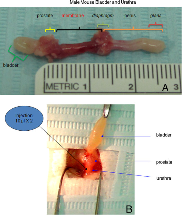
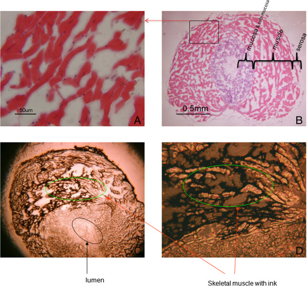
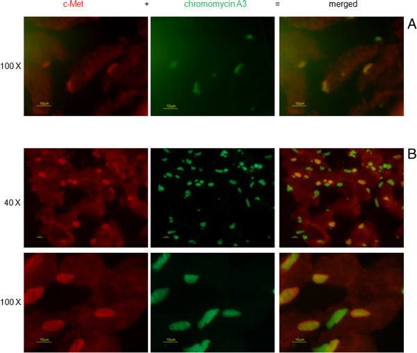
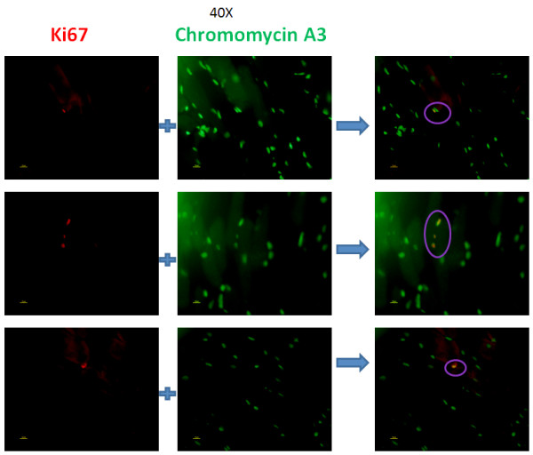
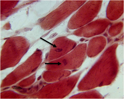
Methods: 20 μl (1 μg/μl) of rIgf-1 was surgically injected directly into the urethral wall of retired male mouse breeders. Mice injected with phosphate buffered saline (PBS) were used as controls. 4 weeks later, urethras were harvested and serially-sectioned through the sphincter for routine hematoxylin-eosin staining as well as immunohistochemical staining with satellite cell specific anti-c-Met antibody and proliferation specific anti-Ki-67 antibody.
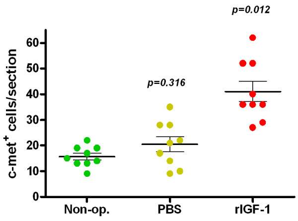
Results: Anti-c-Met antibody positive cells (c-Met+) were identified in the rhabdosphincter. c-Met+ cells increased by 161.8% relative to controls four weeks after rIGF-1 injection. Anti- Ki-67 antibody positive cells were identified and characterized as cells with centrally located nuclei in striated muscle bundles of rIGF-1 treated animals.
Conclusions: Satellite cells in the mouse rhabdosphincter can be activated by rIGF-1 treatment, which subsequently are incorporated into existing skeletal muscle bundles. Using this approach, the rhabdosphincter can be induced to regenerate and potentially strengthen via satellite cell activation and likely improve urinary continence.
Figures
Similar articles
-
rHGF interacts with rIGF-1 to activate the satellite cells in the striated urethral sphincter in rats: a promising treatment for urinary incontinence?Arch Gynecol Obstet. 2018 Dec;298(6):1149-1157. doi: 10.1007/s00404-018-4930-2. Epub 2018 Oct 10. Arch Gynecol Obstet. 2018. PMID: 30306312 Free PMC article.
-
Growth inhibition and apoptosis induction by tumor necrosis factor-α in human urethral rhabdosphincter satellite cells.J Urol. 2010 Jun;183(6):2445-50. doi: 10.1016/j.juro.2010.01.063. Epub 2010 Apr 18. J Urol. 2010. PMID: 20403612
-
Growth mechanism of satellite cells in human urethral rhabdosphincter.Neurourol Urodyn. 2007;26(4):552-561. doi: 10.1002/nau.20369. Neurourol Urodyn. 2007. PMID: 17262837
-
[The striated sphincter of the urethra. 2: Specific methods for studying the striated sphincter of the urethra].J Urol (Paris). 1984;90(8-9):515-27. J Urol (Paris). 1984. PMID: 6398827 Review. French.
-
Insulin-like growth factor 1 and muscle growth: implication for satellite cell proliferation.Proc Nutr Soc. 2004 May;63(2):337-40. doi: 10.1079/PNS2004354. Proc Nutr Soc. 2004. PMID: 15294052 Review.
Cited by
-
Stem cell therapy combined with controlled release of growth factors for the treatment of sphincter dysfunction.Cell Biosci. 2023 Mar 16;13(1):56. doi: 10.1186/s13578-023-01009-3. Cell Biosci. 2023. PMID: 36927578 Free PMC article. Review.
-
rHGF interacts with rIGF-1 to activate the satellite cells in the striated urethral sphincter in rats: a promising treatment for urinary incontinence?Arch Gynecol Obstet. 2018 Dec;298(6):1149-1157. doi: 10.1007/s00404-018-4930-2. Epub 2018 Oct 10. Arch Gynecol Obstet. 2018. PMID: 30306312 Free PMC article.
-
Establishment of new transurethral catheterization methods for male mice.Biol Methods Protoc. 2024 Feb 5;9(1):bpae005. doi: 10.1093/biomethods/bpae005. eCollection 2024. Biol Methods Protoc. 2024. PMID: 38414648 Free PMC article.
-
Controlled release of insulin-like growth factor 1 enhances urethral sphincter function and histological structure in the treatment of female stress urinary incontinence in a rat model.BJU Int. 2018 Feb;121(2):301-312. doi: 10.1111/bju.13985. Epub 2017 Oct 3. BJU Int. 2018. PMID: 28805303 Free PMC article.
-
Histology Atlas of the Developing Mouse Urinary System With Emphasis on Prenatal Days E10.5-E18.5.Toxicol Pathol. 2019 Oct;47(7):865-886. doi: 10.1177/0192623319873871. Epub 2019 Oct 10. Toxicol Pathol. 2019. PMID: 31599209 Free PMC article.
References
Publication types
MeSH terms
Substances
Grants and funding
LinkOut - more resources
Full Text Sources
Other Literature Sources
Miscellaneous
