. 2014 Jul;9(9):1311-26.
doi: 10.2217/nnm.13.137. Epub 2013 Nov 27.

Inhibition of phosphoinositol 3 kinase contributes to nanoparticle-mediated exaggeration of endotoxin-induced leukocyte procoagulant activity

Affiliations

Inhibition of phosphoinositol 3 kinase contributes to nanoparticle-mediated exaggeration of endotoxin-induced leukocyte procoagulant activity

Anna N Ilinskaya et al. Nanomedicine (Lond). 2014 Jul.

Abstract

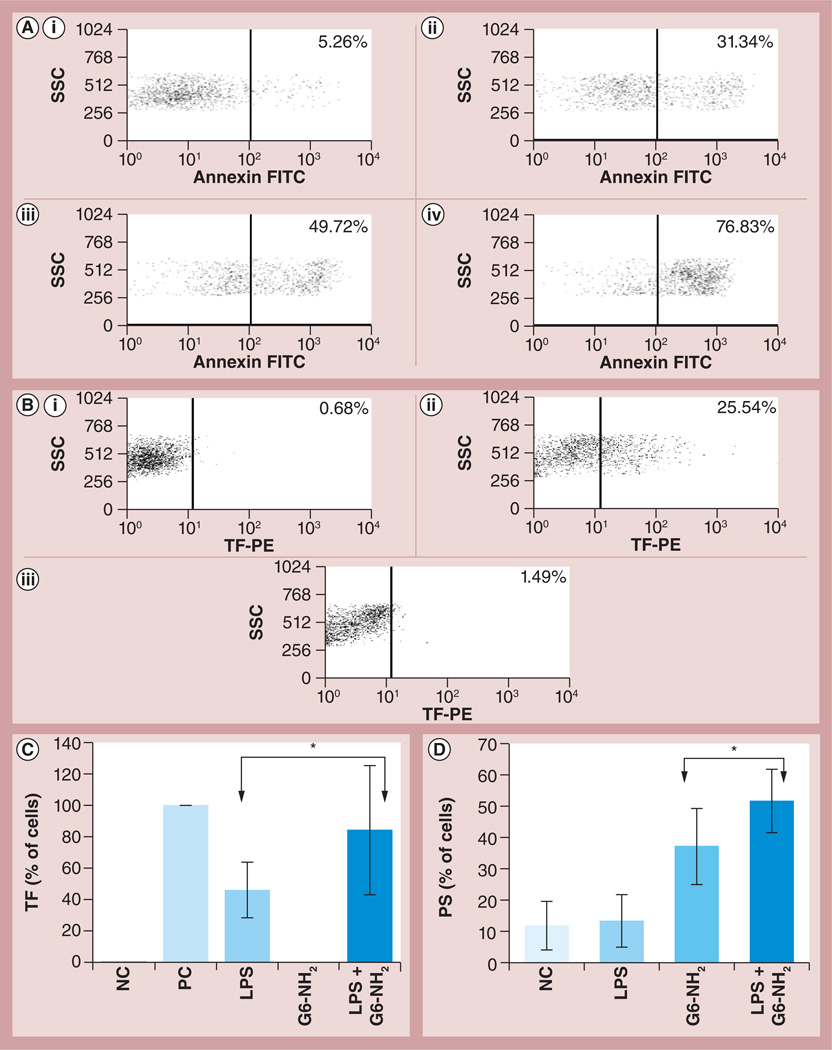
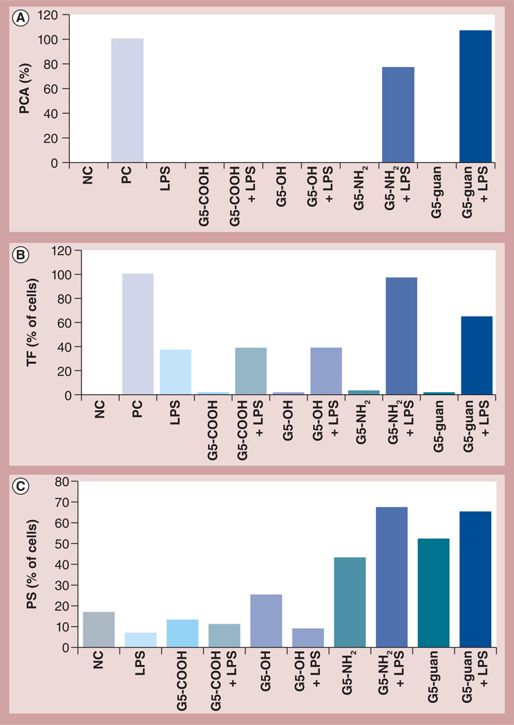
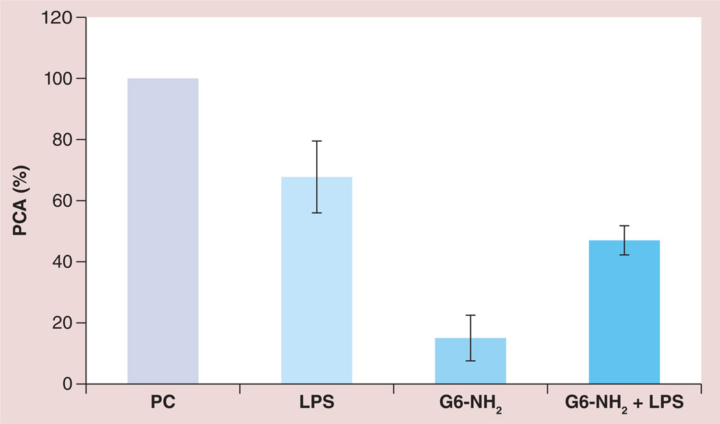
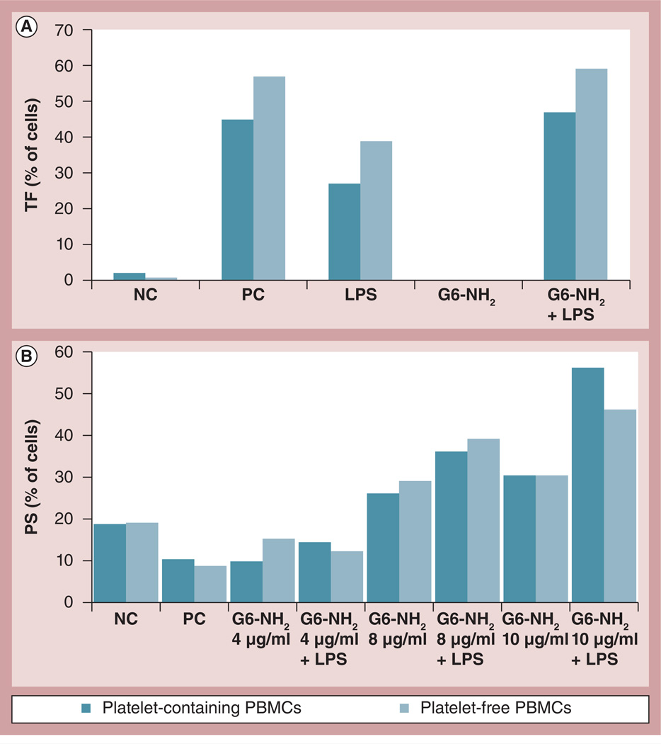
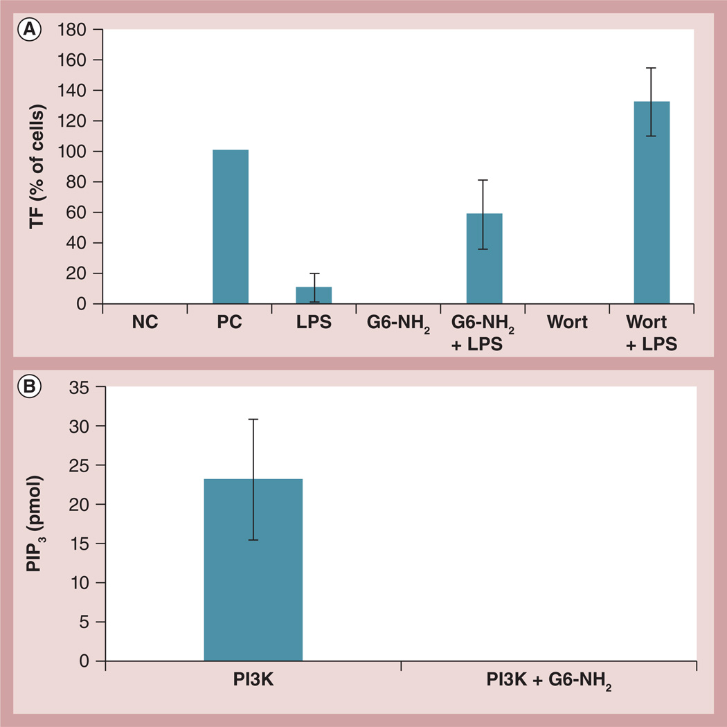
Aim: Disseminated intravascular coagulation is an increasing concern for certain types of engineered nanomaterials. Recent studies have shed some light on the nanoparticle physicochemical properties contributing to this toxicity; however, the mechanisms are poorly understood. Leukocyte procoagulant activity (PCA) is a key factor contributing to the initiation of this toxicity. We have previously reported on the exaggeration of endotoxin-induced PCA by cationic dendrimers. Herein, we report an effort to discern the mechanism.

Materials & methods: Poly(amidoamine) dendrimers with various sizes and surface functionalities were studied in vitro by the recalcification test, flow cytometry and other relevant assays.

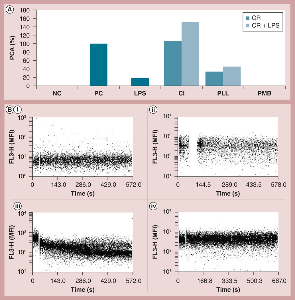
Results & conclusion: Cationic dendrimers exaggerated endotoxin-induced PCA, but their anionic or neutral counterparts did not; the cationic charge prompts this phenomenon, but different cationic surface chemistries do not influence it. Cationic dendrimers and endotoxin differentially affect the PCA complex. The inhibition of phosphoinositol 3 kinase by dendrimers contributes to the exaggeration of the endotoxin-induced PCA.

Keywords: coagulopathy; dendrimer; disseminated intravascular coagulation; leukocyte; nanoparticle; procoagulant activity; thrombosis.


Conflict of interest statement

Financial & competing interests disclosure

The authors have no other relevant affiliations or financial involvement with any organization or entity with a financial interest in or financial conflict with the subject matter or materials discussed in the manuscript apart from those disclosed.

No writing assistance was utilized in the production of this manuscript.

Figures

Figure 1
Figure 1. Flow cytometry analysis of tissue factor expression and phosphatidyl serine presentation on the leukocyte surface in response to endotoxin, dendrimers and the combination thereof
Peripheral blood mononuclear cells from healthy donor volunteers were treated with LPS and G6-NH2 alone or in combination for 24 h. (A) PS presentation on the surface of cells incubated with increasing concentrations of G6-NH2 dendrimers; (i) 4, (ii) 8, (iii) 10 and (iv) 25 µg/ml. (B) TF expression on the surface of cells (i) untreated or incubated with (ii) 1 ng/ml LPS or (iii) 4 µg/ml of G6-NH2 dendrimers. (C) TF expression on the surface of cells treated with 1 ng/ml LPS or 4 µg/ml G6-NH2 dendrimers alone or in combination (LPS + G6-NH2). Shown is the mean value ± standard deviation of percentage-positive cells derived from analysis of ten donor specimens. PC was assigned a 100% value. (D) PS presentation on the surface of cells treated with 1 ng/ml LPS or 10 µg/ml G6-NH2 dendrimers alone or in combination (LPS + G6-NH2). Shown is the mean value ± standard deviation of percentage-positive cells derived from analysis of six donor specimens. *p < 0.05. FITC: Fluorescein isothiocyanate; G6-NH2: Generation 6 amine-terminated polyamidoamine dendrimer; LPS: Lipopolysaccharide; NC: Negative control (cell culture media); PC: Positive control (10 µg/ml LPS); PE: Phycoerythrin; PS: Phosphatidyl serine; SSC: Side scatter; TF: Tissue factor.
Figure 2
Figure 2. Effects of nanoparticle charge and surface chemistry on procoagulant activity
Peripheral blood mononuclear cells from healthy donor volunteers were treated with LPS and G5 dendrimers with different surface groups alone or in combination. (A) Induction of PCA by dendrimers at 25 µg/ml and LPS at 1 ng/ml. PCA was calculated as the percentage of coagulation induced by the PC, LPS at 10 µg/ml. Shown is the mean response (n = 4) for one donor. Similar data were obtained with three additional donors (Supplementary Figure S2). (B) Flow cytometry analysis of TF expression on the surface of monocytes after treatment with various dendrimers at 4 µg/ml (G5-COOH; G5-OH; G5-NH2 and G5-guan), LPS at 1 ng/ml or a combination (G5-COOH + LPS; G5-OH + LPS; G5-NH2 + LPS and G5-guan + LPS). Shown is the percentage of positive cells from one donor relative to the PC. Similar data were obtained with two additional donors (Supplementary Figure S2). PC for this test was LPS (10 µg/ml). (C) Flow cytometry analysis of PS presentation on the surface of monocytes after treatment with various dendrimers at 12 µg/ml (G5-COOH; G5-OH; G5-NH2 and G5-guan), LPS at 1 ng/ml or a combination (G5-COOH + LPS; G5-OH + LPS; G5-NH2 + LPS and G5-guan + LPS). Shown is the percentage of positive (i.e., PS presenting) cells. Similar data were obtained with two additional donors (Supplementary Figure S2). PC for this test was 30 min treatment with formalin (not shown). G5: Generation 5; guan: Guanidine; LPS: Lipopolysaccharide; NC: Negative control; PC: Positive control; PCA: Procoagulant activity; PS: Phosphatidyl serine; TF: Tissue factor.
Figure 3
Figure 3. Interaction between lipopolysaccharide and dendrimers are not responsible for the enhancement of lipopolysaccharide-induced procoagulant activity by the dendrimers
Peripheral blood mononuclear cells from healthy donor volunteers were treated with 1 ng/ml of LPS and 25 µg/ml G6-NH2 poly(amidoamine) dendrimers under the following conditions: LPS and G6-NH2 were added to cells concurrently (LPS + G6); cells were treated with G6-NH2 for 6 h, then washed with phosphate-buffered saline and treated with LPS for an additional 12–18 h (G6-6 h-LPS); or cells were treated with LPS for 6 h, then washed with phosphate-buffered saline and treated with G6-NH2 for an additional 12–18 h (LPS-6 h-G6). PCA was assessed 24 h after addition of the first treatment. PCA was calculated as a percentage relative to that induced by the positive control LPS at 10 µg/ml. G6: Generation 6; LPS: Lipopolysaccharide; PCA: Procoagulant activity.
Figure 4
Figure 4. Understanding the role of cationic charge and Ca2+ influx in the enhancement of lipopolysaccharide-induced procoagulant activity
(A) Peripheral blood mononuclear cells from healthy donor volunteers were treated with different cationic reagents: poly-l-lysine at 25 µg/ml (~1 µM), Ca+ ionophore at 1 µM; or PMB at 10 µg/ml (~8 µM). Peripheral blood mononuclear cells were treated with the CR alone or in combination with 1 ng/ml of LPS (CR + LPS) for 24 h. To avoid possible neutralizing effects of PMB on LPS, the cells in the PMB + LPS group were first treated with LPS for 6 h, washed with PBS, then challenged with PMB for an additional 12–18 h. PCA activity was measured 24 h after the first treatment. PCA was calculated as a percentage relative to that induced by the positive control LPS at 10 µg/ml. Similar results were obtained with two additional donors (Supplementary Figure S3). (B) Flow cytometry analysis of Ca2+ influx. Unloaded cells were treated with (i) Ca2+ ionophore as control; (ii) peripheral blood mononuclear cells loaded with Fura Red® (Molecular Probe, OR, USA) were untreated or (iii) treated with 1 µM Ca2+ ionophore or (iv) 25 µg/ml generation 6 amine-terminated polyamidoamine dendrimer. The fluorescence emission of Fura Red decreases with the increase of the intracellular Ca2+ (see manufacturer’s instructions for details). CR: Cationic reagent; FL3-H: Fluorescent channel 3; LPS: Lipopolysaccharide; MFI: Mean fluorescent intensity; PCA: Procoagulant activity; PLL: Poly-l-lysine; PMB: Polymyxin B.
Figure 5
Figure 5. Procoagulant activity of MM-6 cells in response to lipopolysaccharide, cationic dendrimers and the combination thereof
MM-6 cells were treated with 1 ng/ml LPS, 25 µg/ml amine-terminated generation 6 poly(amidoamine) dendrimers, or a combination of both LPS and G6-NH2 (G6-NH2 + LPS). PCA is calculated as a percentage of that induced by the positive control. Shown is the mean response ± standard deviation of three independent experiments (n = 3). G6-NH2: Generation 6 amine-terminated polyamidoamine dendrimer; LPS: Lipopolysaccharide; PC: Positive control (lipopolysaccharide; 10 µg/ml); PCA: Procoagulant activity.
Figure 6
Figure 6. Role of platelets in enhancement of endotoxin-induced procoagulant activity by amine-terminated dendrimers
Platelet-containing and platelet-free PBMCs from healthy donor volunteers were treated with 1 ng/ml of LPS or G6-NH2 alone (LPS and G6-NH2, respectively) or in combination (LPS + G6-NH2) for 24 h. (A) TF expression and (B) phosphatidyl serine presentation were analyzed by flow cytometry. TF was assessed using 4 µg/ml G6-NH2. Phosphatidyl serine was assessed using 4, 8 and 10 µg/ml G6-NH2. Shown is the mean percentage of positive cells. Shown are data from a single donor. Data from additional donors are provided in Supplementary Figure S5. LPS: Lipopolysaccharide; G6-NH2: Generation 6 amine-terminated polyamidoamine dendrimer; NC: Negative control; PBMC: Peripheral blood mononuclear cell; PC: Positive control; TF: Tissue factor.
Figure 7
Figure 7. Probing the role of phosphoinositol 3 kinase in the enhancement of lipopolysaccharide-induced procoagulant activity by cationic dendrimers
(A) Peripheral blood mononuclear cells from healthy donor volunteers were treated for 24 h with 1 ng/ml of LPS, 4 µg/ml of G6-NH2, or the combination (LPS + G6-NH2). Cells were also treated with 100 nM Wort alone or in combination with LPS (Wort + LPS). Tissue factor expression on monocyte surfaces was measured by flow cytometry. Shown is the mean percentage of positive cells relative to the positive control. Media was used as the NC and 10 µg/ml of LPS was used as the PC. (B) Recombinant human PI3K was immunoprecipitated with anti-p85 antibody followed by several washes; the beads with isolated PI3K were then incubated with 12.5 µg/ml G6-NH2. After eight washes to remove excess dendrimer, the kinase activity was assessed by incubation with phosphatidylinositol (4,5)-biphosphate substrate. Shown is the mean amount of PIP3 generated from the kinase reaction ± standard deviation (n = 3). G6-NH2: Generation 6 amine-terminated polyamidoamine dendrimer; LPS: Lipopolysaccharide; NC: Negative control; PC: Positive control; PI3K: Phosphoinositol 3 kinase; PIP3: Phosphatidylinositol (3,4,5)-triphosphate; Wort: Wortmannin.

Similar articles

Cited by

References

    1. Levi M. Disseminated intravascular coagulation. Crit. Care Med. 2007;35(9):2191–2195. - PubMed
    1. Levi M, Schultz M, van der Poll T. Disseminated intravascular coagulation in infectious disease. Semin. Thromb. Hemost. 2010;36(4):367–377. - PubMed
    1. Franchini M, Di Minno MN, Coppola A. Disseminated intravascular coagulation in hematologic malignancies. Semin. Thromb. Hemost. 2010;36(4):388–403. - PubMed
    1. Lippi G, Cervellin G. Disseminated intravascular coagulation in trauma injuries. Semin. Thromb. Hemost. 2010;36(4):378–387. - PubMed
    1. Montagnana M, Franchi M, Danese E, Gotsch F, Guidi GC. Disseminated intravascular coagulation in obstetric and gynecologic disorders. Semin. Thromb. Hemost. 2010;36(4):404–418. - PubMed

Publication types

MeSH terms

LinkOut - more resources